Quick Facts
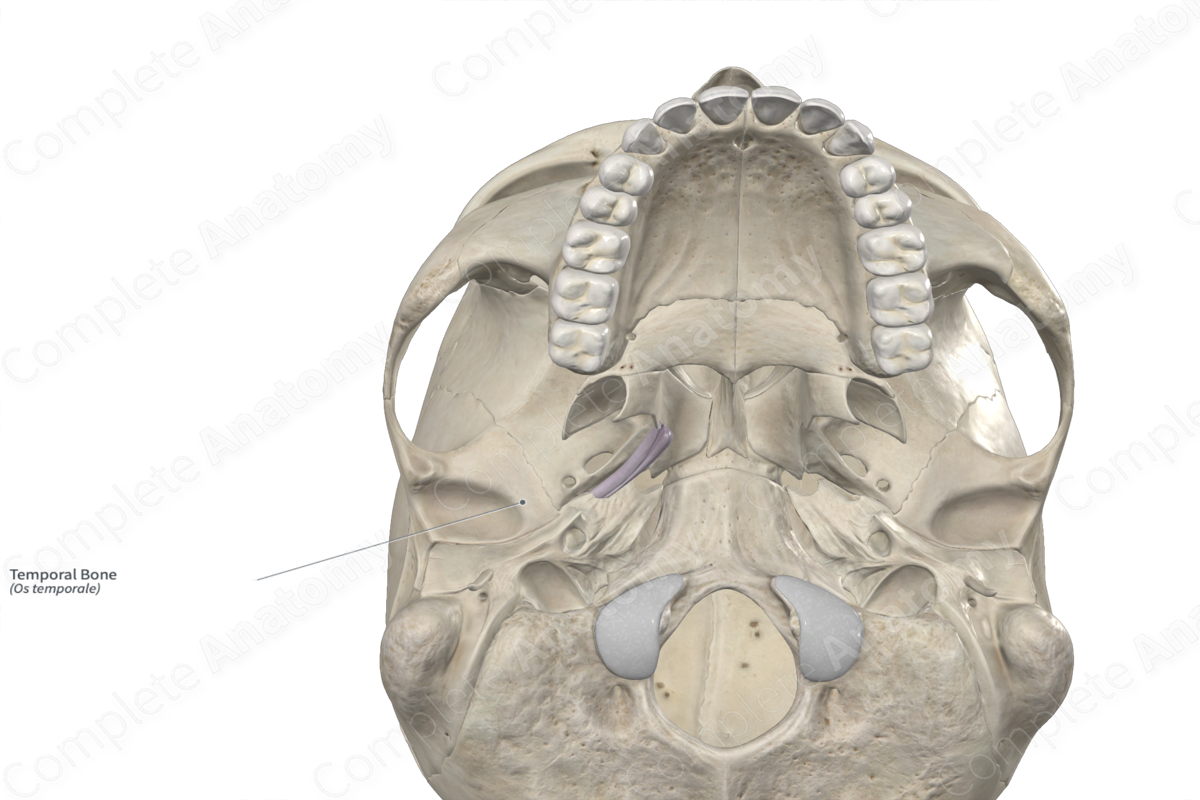
Location: Neurocranium.
Bone Type: Irregular bone.
Key Features: Squamous, petrous, tympanic, and mastoid parts, styloid process, mandibular fossa, and zygomatic and mastoid processes.
Articulates With: Parietal, occipital, sphenoid, and zygomatic bones.
Arterial Supply: Middle meningeal, posterior auricular, and stylomastoid arteries.
Key Features & Anatomical Relations
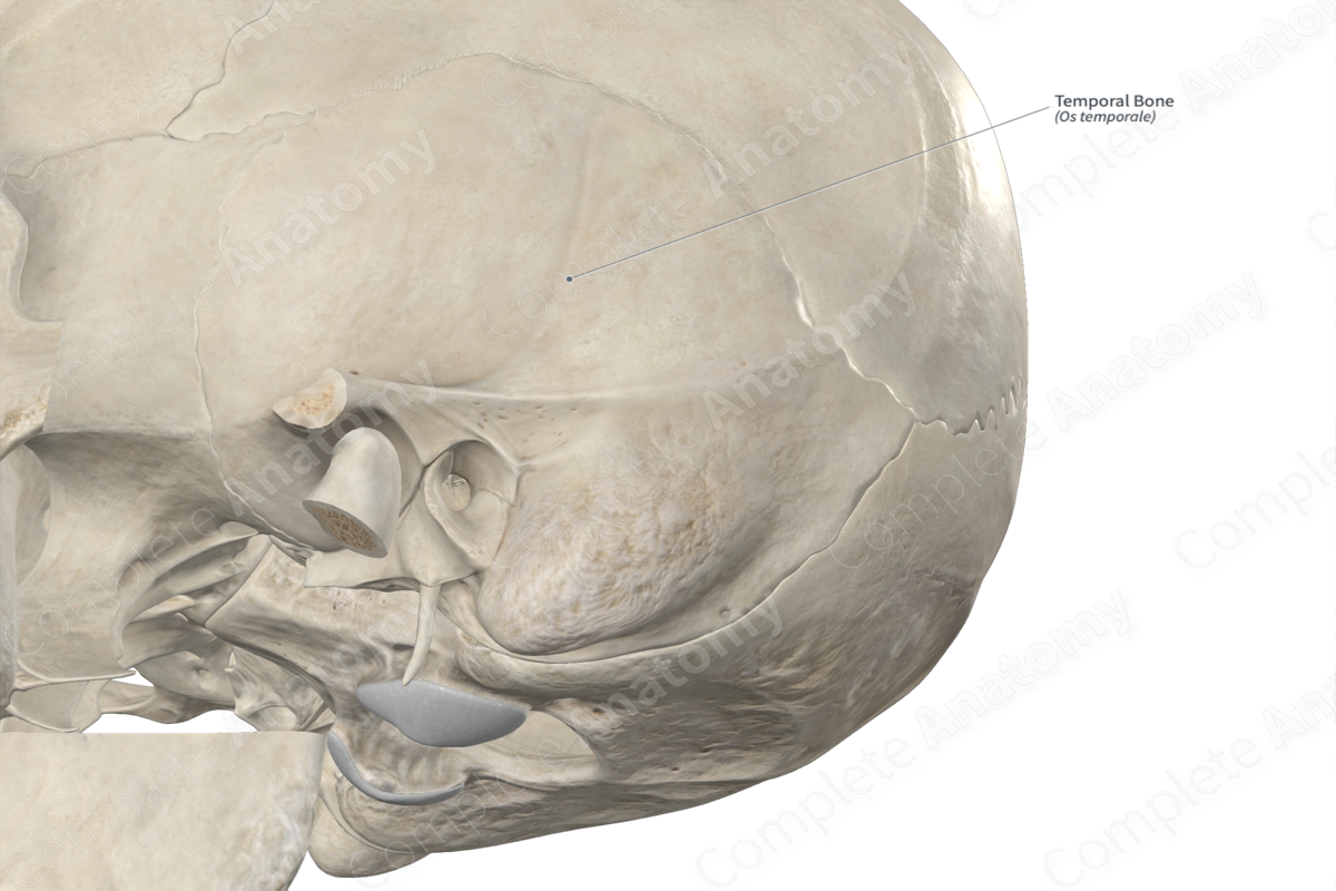
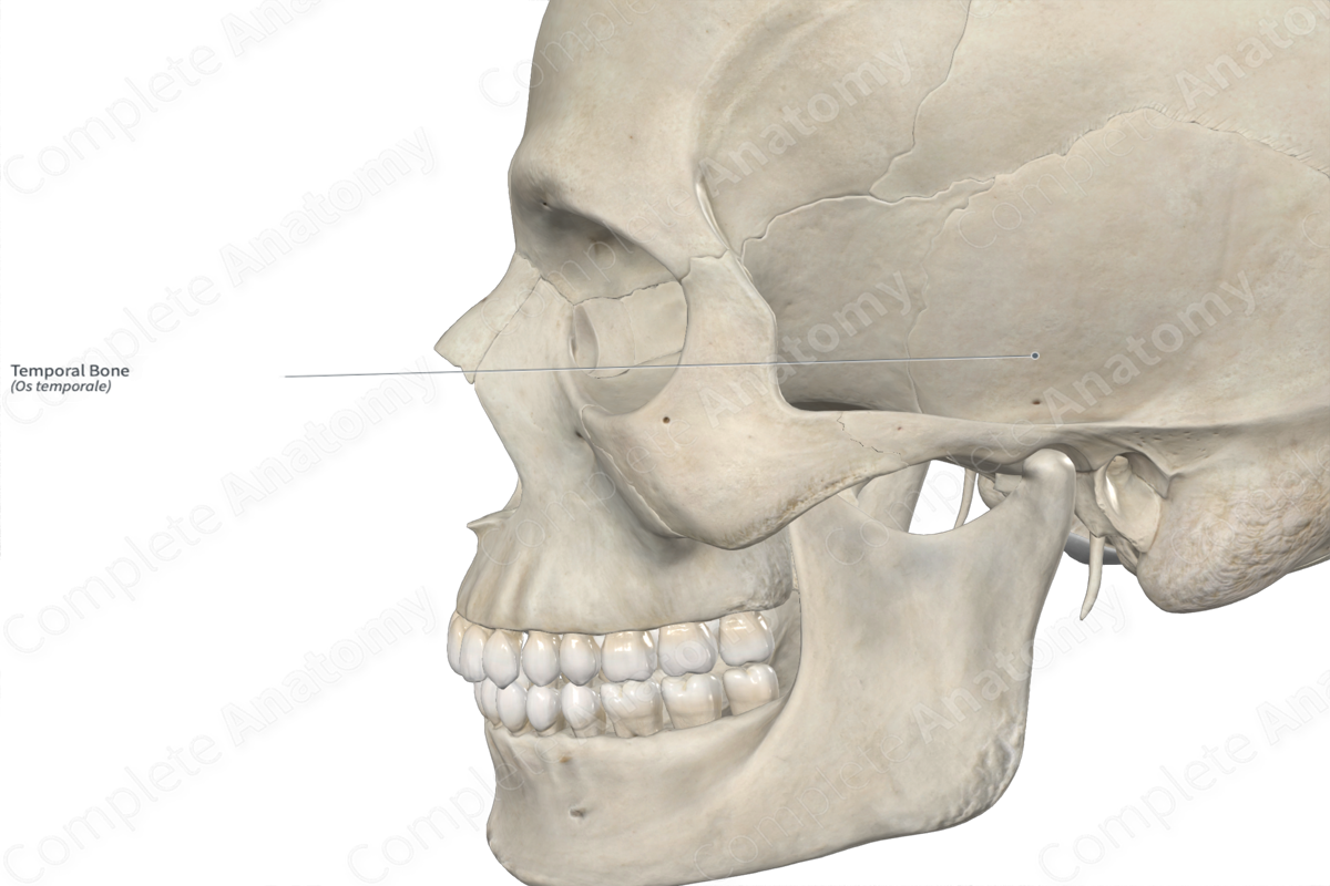
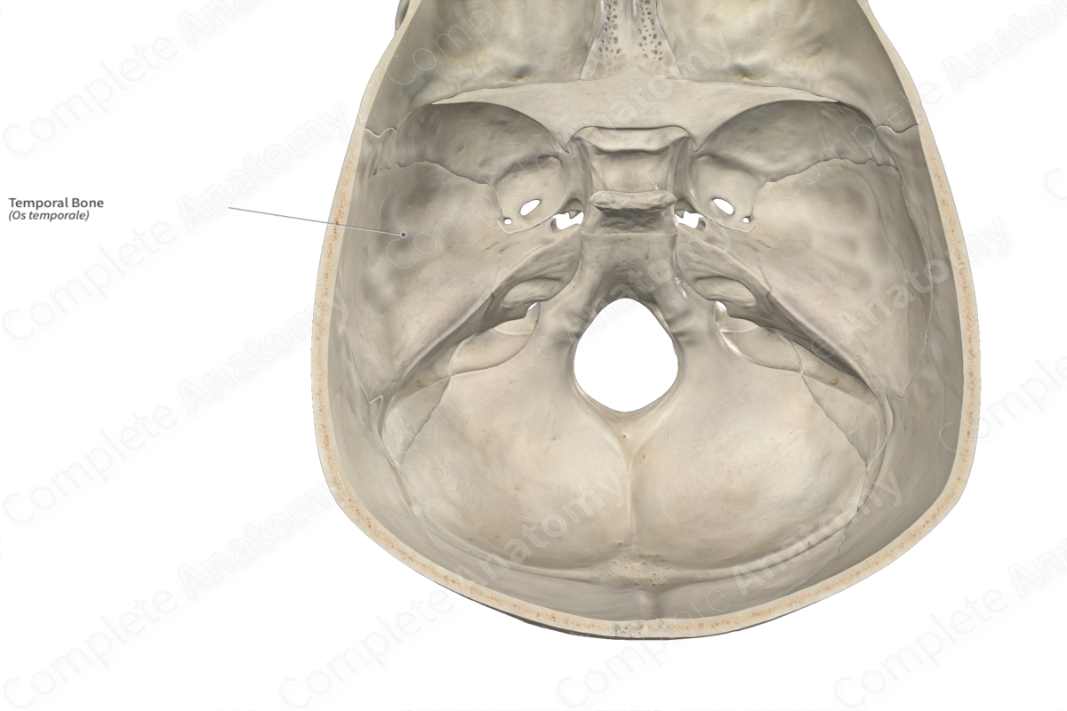
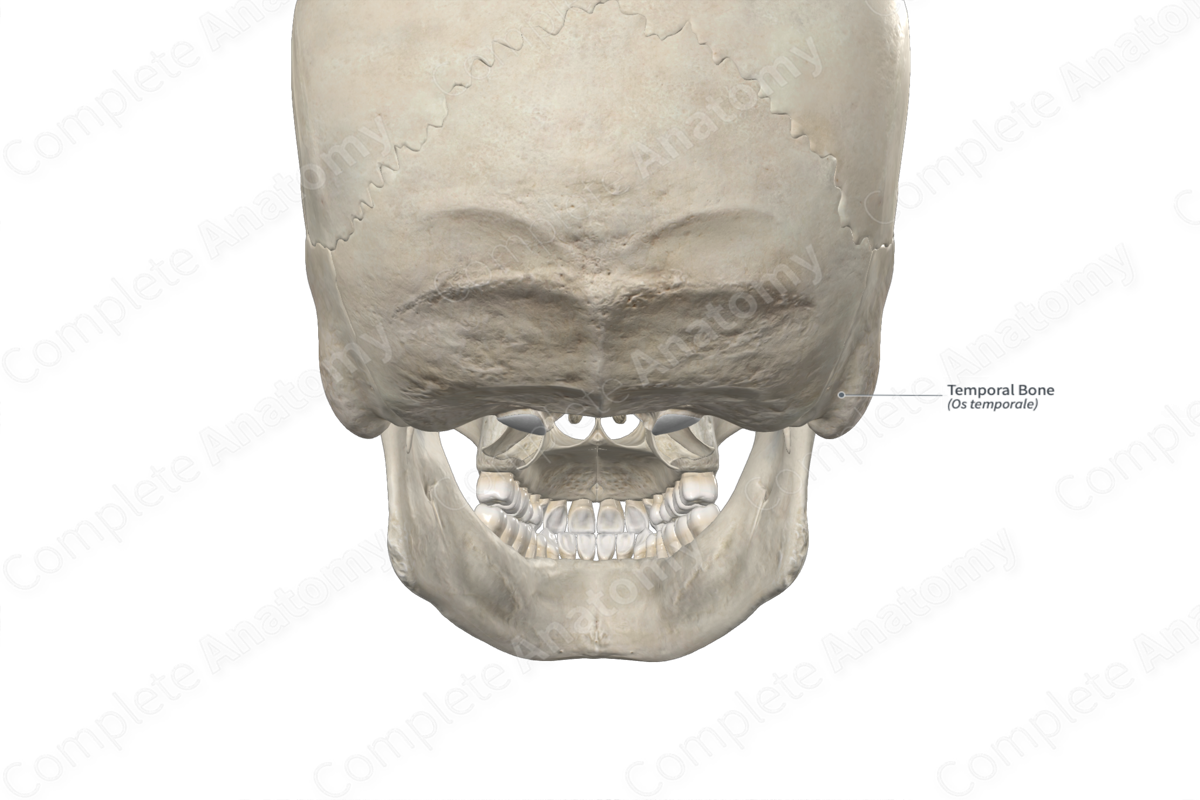
The temporal bones are a pair of large bones found along the inferolateral aspects of the cranium. They are classified as irregular bones, contain the middle ear and internal ear, and contribute to the formation of the neurocranium. Each temporal bone includes the following bony features:
- parts: Squamous, petrous, tympanic, and mastoid parts, and styloid process;
- surfaces: anterior, posterior, and inferior surfaces of petrous part, temporal and cerebral surfaces of squamous part, and internal and external surfaces of mastoid part;
- landmarks: mastoid process, tegmen tympani, arcuate eminence, jugular fossa, supramastoid crest, and postglenoid tubercle.
More information regarding these bony features can be found in the Parts, Surfaces and Landmarks tabs for this bone.
On its corresponding side, each temporal bone is located:
- anterolateral to the occipital bone;
- posterior to the sphenoid and zygomatic bones;
- posterosuperior to the mandible;
- inferior to the parietal bone.
Each temporal bone articulates with the:
- occipital bone at the occipitomastoid suture and petrooccipital synchondrosis;
- parietal bone at the squamous and parietomastoid sutures;
- sphenoid bone at the sphenosquamous suture and sphenopetrosal synchondrosis;
- zygomatic bone at the temporozygomatic suture;
- mandible at the temporomandibular joints.
Ossification
Ossification of each temporal bone occurs at many ossification centers, these are found in the:
- squamous part, with one center found in it, which appears in utero during the second month;
- tympanic part, with one to four centers found in it, which appear in utero during the third month;
- petrous and mastoid parts, with many centers found in these, which appear in utero after the fifth month;
- styloid process, with two centers found in it, which appear around the time of birth;
The ossification centers for the tympanic and squamous parts fuse with each other before birth. The petrous and mastoid parts and the styloid process fuse with the rest of the temporal bone during the first year after birth (Standring, 2020).
Variations
In some individuals:
- the styloid process may be present in a more elongated form;
- a foramen tympanicum (foramen of Hüschke) may be present along the bony part of the external acoustic meatus (Tubbs, Shoja and Loukas, 2016);
- the frontal and temporal bones meet each other and articulate, resulting in the parietal and sphenoid bones not meeting and articulating.
The temporal bone also displays sexual dimorphism, where the mastoid processes and temporal lines tend to be less prominent and more gracile in females than in males (Standring, 2020; White and Folkens, 2005).
Surface Anatomy
The following bony features of each temporal bone are relevant to surface anatomy:
- the mastoid process is palpable posterior to the external ear;
- the zygomatic arch, which is formed by both the zygomatic process of temporal bone and the temporal process of zygomatic bone, is palpable.
List of Clinical Correlates
- Fracture of temporal bone
- Osteitis deformans (Paget disease of bone)
- Osteomyelitis of temporal bone
- Mastoiditis of temporal bone
- Craniosynostosis
- Zygomatic arch dysplasia
References
Standring, S. (2020) Gray's Anatomy: The Anatomical Basis of Clinical Practice. 42nd edn.: Elsevier Health Sciences.
Tubbs, R. S., Shoja, M. M. and Loukas, M. (2016) Bergman's Comprehensive Encyclopedia of Human Anatomic Variation. Wiley.
White, T. D. and Folkens, P. A. (2005) The Human Bone Manual. Elsevier Science.
Learn more about this topic from other Elsevier products

