Quick Facts
Location: Neurocranium.
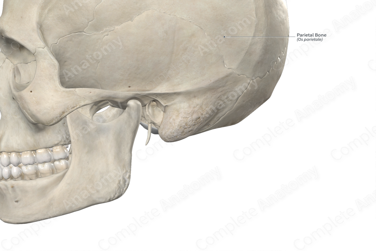
Bone Type: Flat bone.
Key Features: Frontal, occipital, sphenoidal, and mastoid angles, internal and external surfaces, parietal eminence, and superior and inferior temporal lines.
Articulates With: Opposite parietal bone, frontal, occipital, temporal, and sphenoid bones.
Arterial Supply: Middle meningeal artery.
Key Features & Anatomical Relations
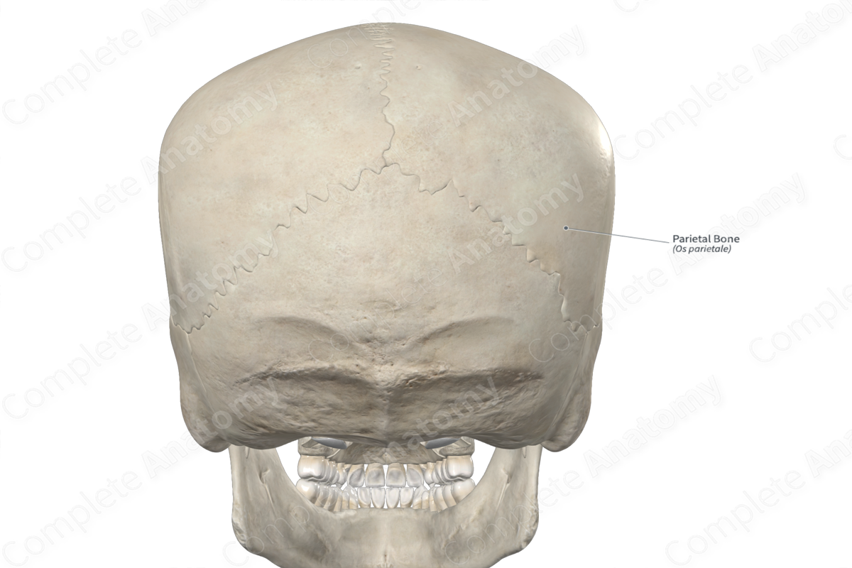
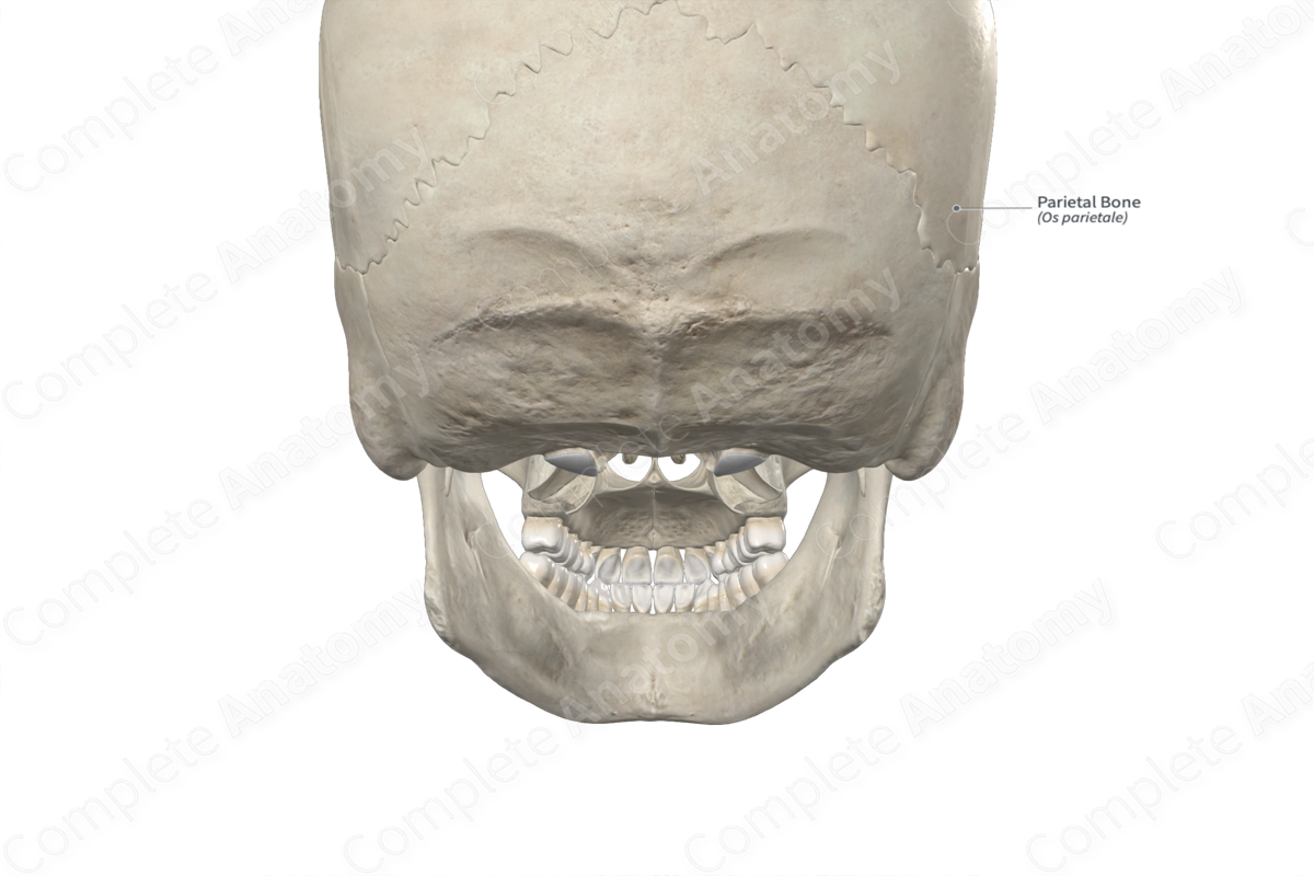
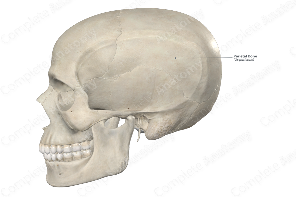
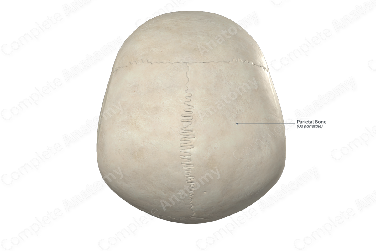
The parietal bones are a pair of large, quadrilateral bones found along the superolateral aspects of the cranium. They are classified as flat bones and contribute to the formation of the neurocranium. Each parietal bone includes the following bony features:
- parts: frontal, occipital, sphenoidal and mastoid angles;
- surfaces: internal and external surfaces, and sagittal, occipital, frontal and squamosal borders;
- landmarks: superior and inferior temporal lines, parietal eminence, and grooves for the sigmoid and superior sagittal sinuses.
More information regarding these bony features can be found in the Parts, Surfaces and Landmarks tabs for this bone.
On its corresponding side, each parietal bone is located:
- anterosuperior to the occipital bone;
- posterior to the frontal bone;
- superior to the temporal and sphenoid bones.
Each parietal bone articulates with the:
- opposite parietal bone at the sagittal suture;
- frontal bone at the coronal suture;
- occipital bone at the lambdoid suture;
- temporal bone at the squamous and parietomastoid sutures;
- sphenoid bone at the sphenoparietal suture.
Ossification
Ossification of a parietal bone occurs at two ossification centers. These are found near the parietal eminence and appear in utero during the second month. They fuse with each other in utero (Standring, 2016).
Variations
In some individuals:
- an accessory intraparietal suture may be present (Tubbs, Shoja and Loukas, 2016);
- the frontal and temporal bones meet each other and articulate, resulting in the parietal and sphenoid bones not meeting and articulating.
Surface Anatomy
Regarding surface anatomy, the parietal eminence and temporal lines of each parietal bone can be palpated.
List of Clinical Correlates
- Fracture of parietal bone
- Enlarged parietal foramina
- Craniosynostosis
References
Standring, S. (2016) Gray's Anatomy: The Anatomical Basis of Clinical Practice. Gray's Anatomy Series 41st edn.: Elsevier Limited.
Tubbs, R. S., Shoja, M. M. and Loukas, M. (2016) Bergman's Comprehensive Encyclopedia of Human Anatomic Variation. Wiley.
Learn more about this topic from other Elsevier products

