Quick Facts
Location: Neurocranium.
Bone Type: Flat bone.
Key Features: Squamous, basilar, and lateral parts, cerebral and cerebellar fossae, superior and inferior nuchal lines, and internal and external occipital protuberances.
Articulates With: Parietal, temporal, and sphenoid bones, and atlas.
Arterial Supply: Middle meningeal artery.
Related parts of the anatomy
Key Features & Anatomical Relations
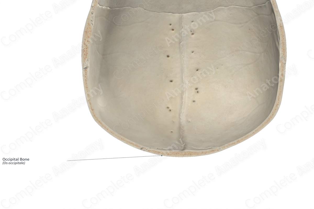
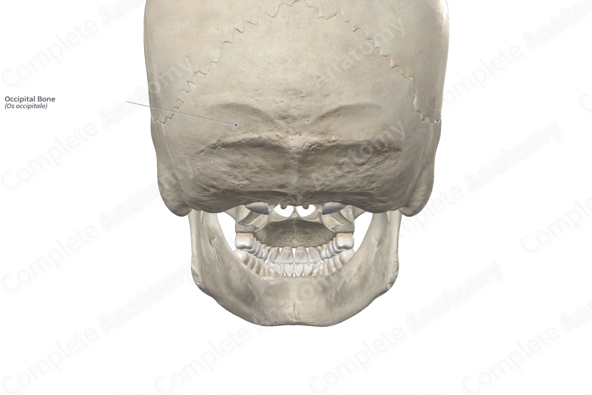
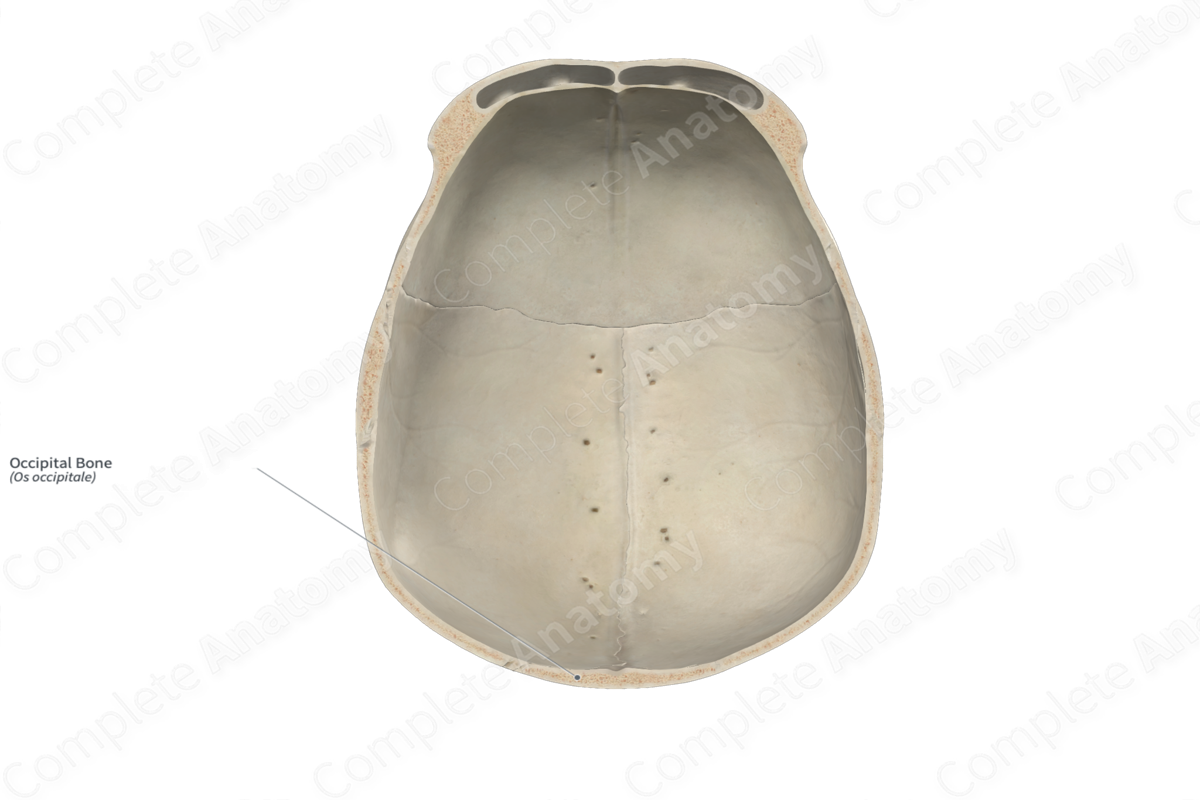
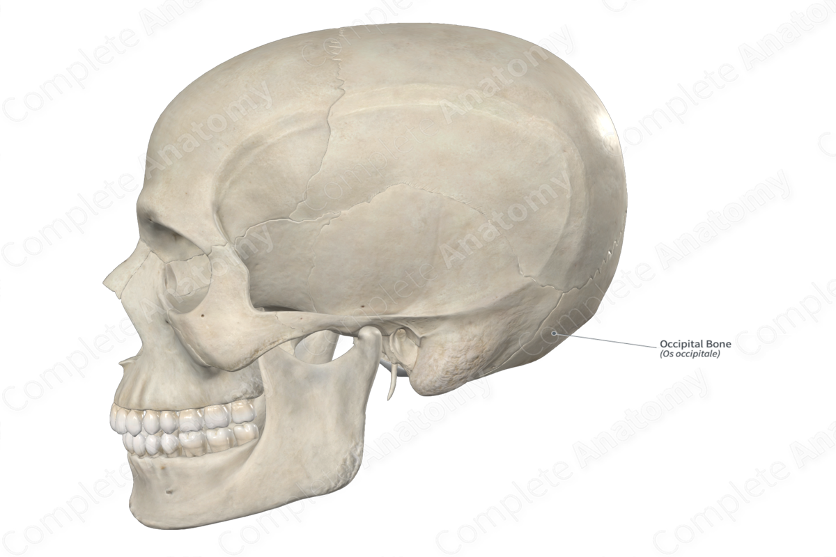
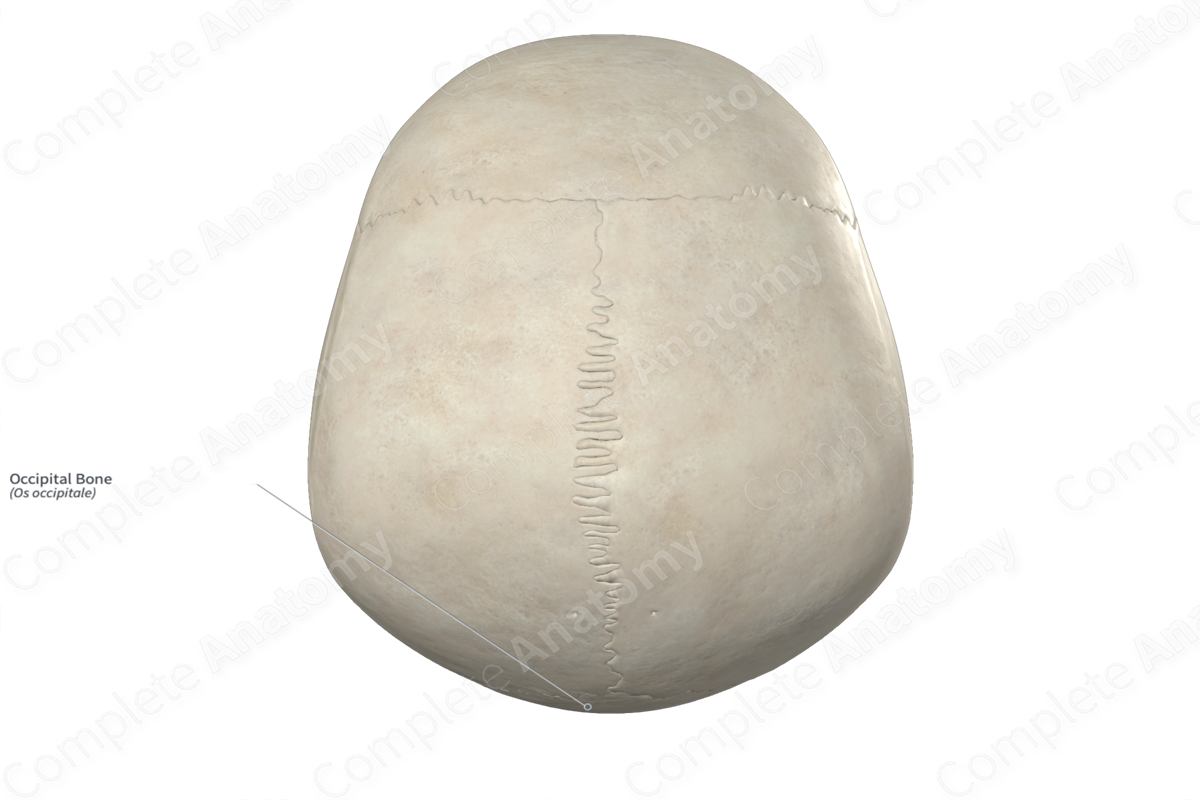
The occipital bone is the single, large bone found along the posteroinferior aspect of the cranium. It is classified as a flat bone, forms the foramen magnum and contributes to the formation of the neurocranium. The occipital bone includes the following bony features:
- parts: squamous, basilar, and lateral parts;
- surfaces: cerebral and cerebellar fossae, superior and lateral angles, and mastoid and lambdoid borders;
- landmarks: supreme, superior and inferior nuchal lines, internal and external occipital protuberances, occipital condyles, and internal and external occipital crests.
More information regarding these bony features can be found in the Parts, Surfaces and Landmarks tabs for this bone.
The occipital bone is located:
- posteroinferior to the parietal bones;
- posteromedial to the temporal bones;
- posterior to the sphenoid bone;
- superior to the atlas (first cervical vertebra).
It articulates with the:
- parietal bones at the lambdoid suture;
- temporal bones at the occipitomastoid sutures and the petrooccipital synchondroses;
- sphenoid bone at the sphenooccipital synchondroses;
- atlas at the atlantooccipital joints.
Ossification
Ossification of the occipital bone occurs at nine ossification centers, these are found in the:
- squamous part, with four centers found here, which appear in utero during the second month;
- basilar part, with one center found here, which appears in utero during the second month;
- right and left lateral parts, with two centers found in each, which appear in utero during the second month.
At birth, the squamous, basilar and lateral parts of the occipital bone remain unfused. The squamous and lateral parts fuse with each other during the second year, while the lateral and basilar parts fuse with each other during the third to fourth years (Standring, 2020).
Variations
In some individuals:
- a third occipital condyle may be present;
- the occipital bone may be fused with the atlas, known as atlantooccipital assimilation;
- the superior portion of the squamous part of occipital bone may fail to fuse with the rest of the bone, resulting in the presence of an interparietal bone (Inca bone) (Tubbs, Shoja and Loukas, 2016).
The occipital bone also displays sexual dimorphism, where the nuchal lines and external occipital protuberance tend to be heavier in males than females (Standring, 2020; White and Folkens, 2005).
Surface Anatomy
Regarding surface anatomy, the external occipital protuberance and superior nuchal lines of the occipital bone can be palpated.
List of Clinical Correlates
- Fracture of occipital bone
- Occipital horn syndrome
- Craniosynostosis
References
Standring, S. (2020) Gray's Anatomy: The Anatomical Basis of Clinical Practice. 42nd edn.: Elsevier Health Sciences.
Tubbs, R. S., Shoja, M. M. and Loukas, M. (2016) Bergman's Comprehensive Encyclopedia of Human Anatomic Variation. Wiley.
White, T. D. and Folkens, P. A. (2005) The Human Bone Manual. Elsevier Science.
Learn more about this topic from other Elsevier products

