Quick Facts
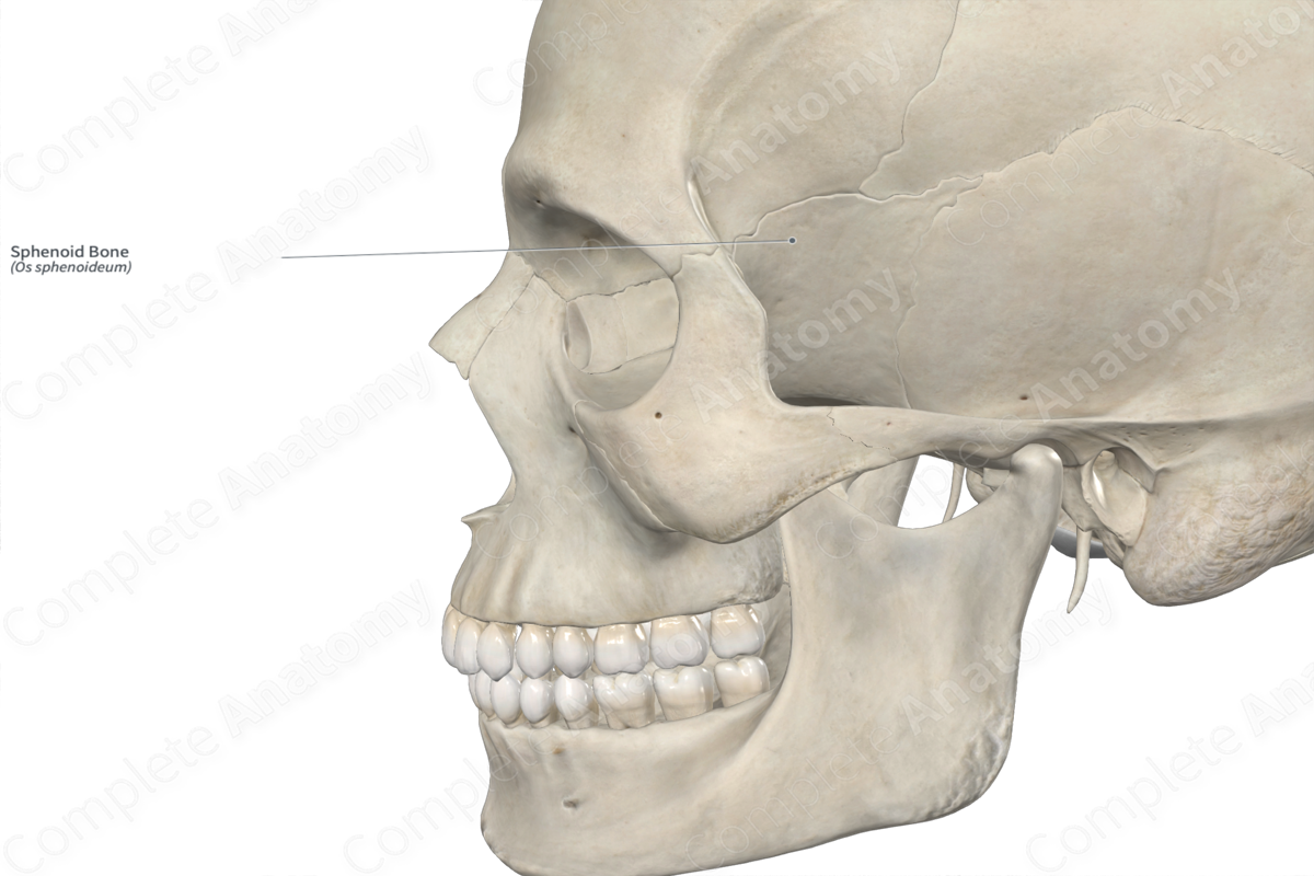
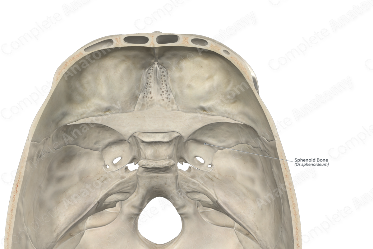
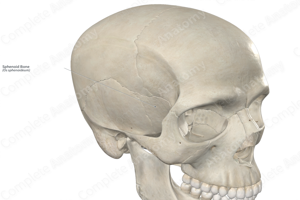
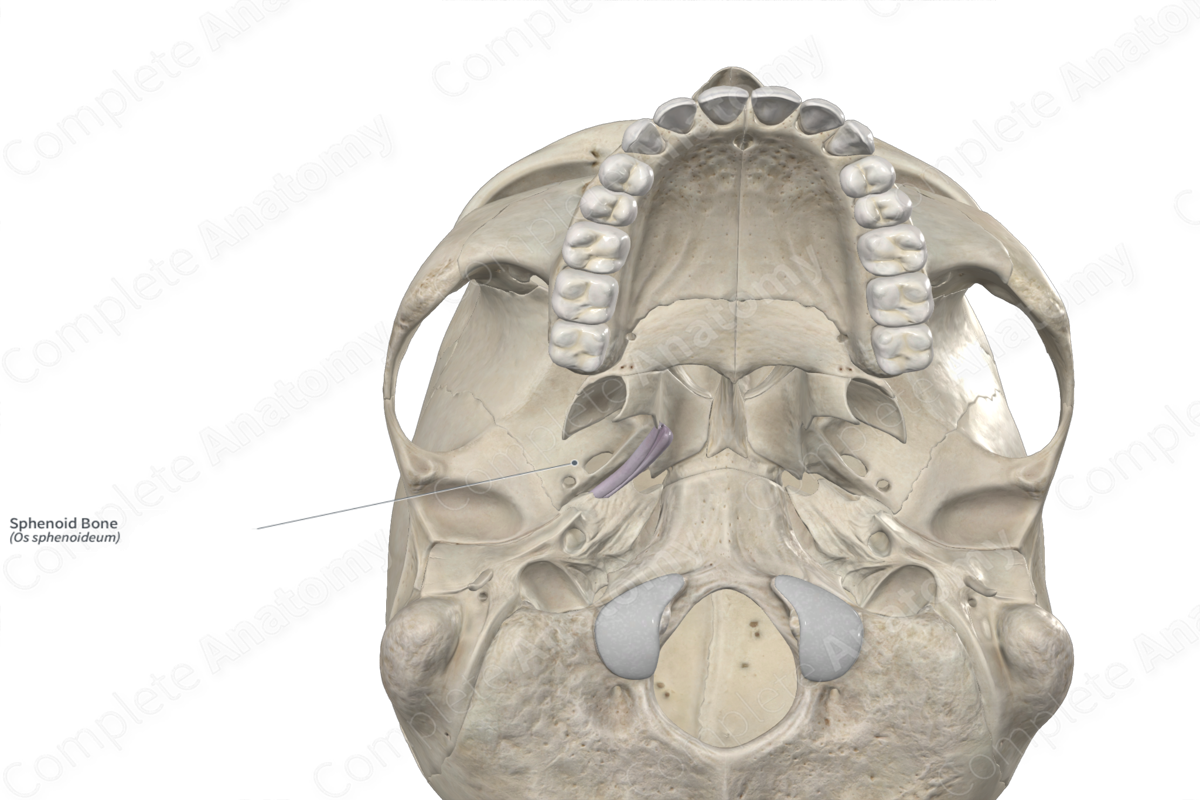
Location: Neurocranium.
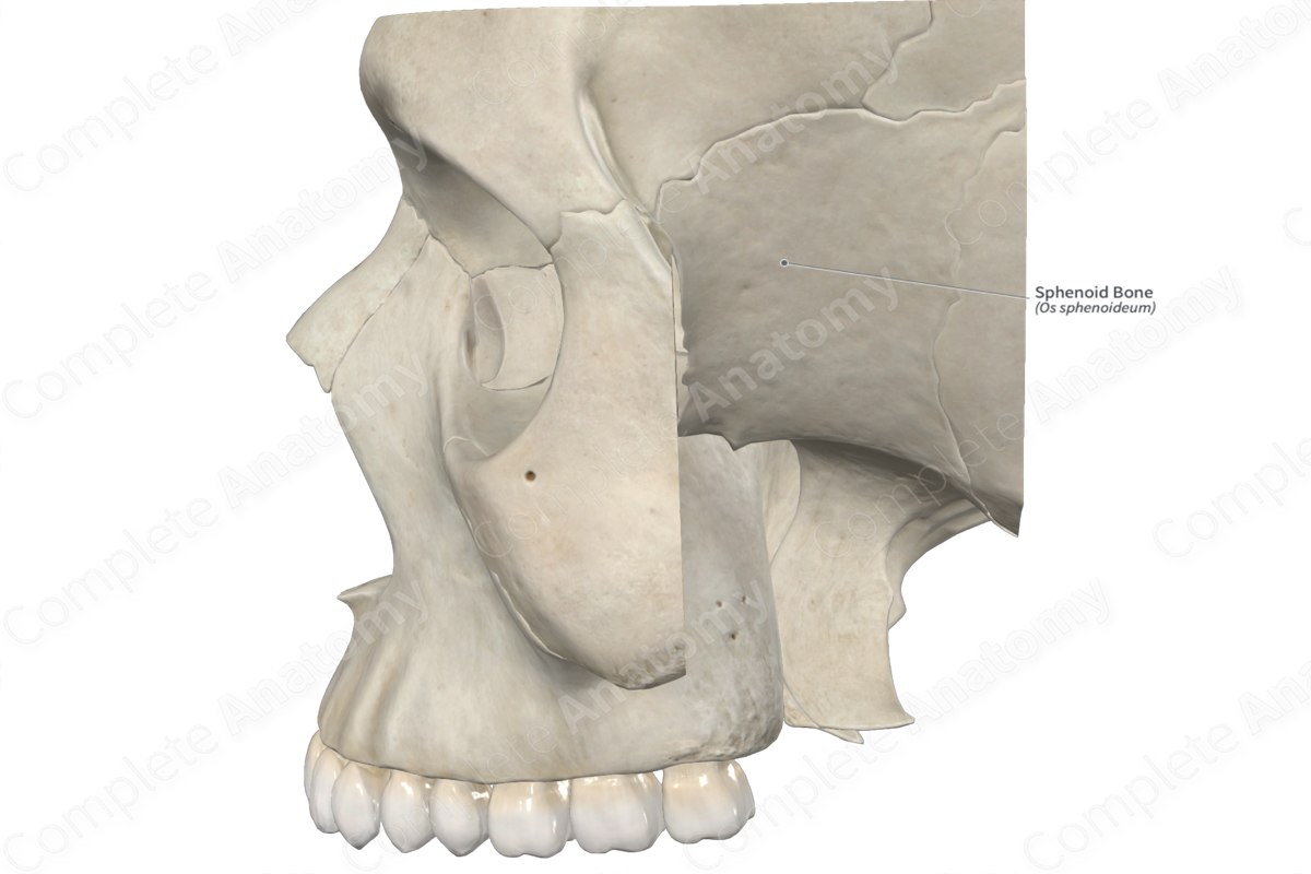
Bone Type: Irregular bone.
Key Features: Body, greater and lesser wings, medial and lateral plates of pterygoid process, sella turcica, and anterior and posterior clinoid processes.
Articulates With: Frontal, parietal, temporal, occipital, zygomatic, ethmoid, and palatine bones, and vomer.
Arterial Supply: Middle meningeal and internal carotid arteries.
Key Features & Anatomical Relations
The sphenoid bone is a single, large bone that is located centrally within the cranium. It is classified as an irregular bone and contributes to the formation of the neurocranium. The sphenoid bone consists of right and left sphenoidal sinuses and includes the following bony features:
- parts: body, greater and lesser wings, and pterygoid processes;
- surfaces: anterior, posterior, and inferior surfaces of body, cerebral, temporal, infratemporal, maxillary, and orbital surfaces of greater wings, and superior and inferior surfaces of lesser wings;
- landmarks: anterior and posterior clinoid processes, chiasmatic sulcus, tuberculum sellae, dorsum sellae, and carotid sulci.
More information regarding these bony features can be found in the Parts, Surfaces and Landmarks tabs for this bone.
The sphenoid bone is located:
- anterior to the temporal and occipital bones;
- posterior to the maxillae and the zygomatic, palatine and ethmoid bones;
- inferior to the frontal and parietal bones.
It articulates with the:
- frontal bone at the sphenofrontal suture;
- parietal bones at the sphenoparietal sutures;
- temporal bones at the sphenosquamous sutures and sphenopetrosal synchondroses;
- occipital bone at the sphenooccipital synchondroses;
- zygomatic bones at the sphenozygomatic sutures;
- ethmoid bone at the sphenoethmoidal suture;
- palatine bones;
- vomer at the sphenovomerine suture.
Ossification
Ossification of the sphenoid bone occurs at many ossification centers, these are found in the:
- body, which appears in utero during the third to fifth months;
- greater and lesser wings, which appear in utero during the third month.
The ossification centers for the body of sphenoid bone fuse with each other in utero. The lesser wings fuse with the body in utero, while the greater wings fuse with the body during the first year after birth (Standring, 2016).
Variations
In some individuals:
- the sella turcica may be absent;
- the sphenoidal sinuses may be present in different sizes and shapes;
- a sphenoidal emissary foramen (foramen of Vesalius) may be present on the greater wing of sphenoid bone (Tubbs, Shoja and Loukas, 2016);
- the frontal and temporal bones meet each other and articulate, resulting in the parietal and sphenoid bones not meeting and articulating.
List of Clinical Correlates
- Fracture of sphenoid bone
- Osteomyelitis of sphenoid bone
- Craniosynostosis
References
Standring, S. (2016) Gray's Anatomy: The Anatomical Basis of Clinical Practice. Gray's Anatomy Series 41st edn.: Elsevier Limited.
Tubbs, R. S., Shoja, M. M. and Loukas, M. (2016) Bergman's Comprehensive Encyclopedia of Human Anatomic Variation. Wiley.
Learn more about this topic from other Elsevier products

