Quick Facts
Location: Neurocranium.
Bone Type: Flat bone.
Key Features: Squamous, orbital and nasal parts, supraorbital margin, superciliary arch, fossae for lacrimal glands, and zygomatic processes.
Articulates With: Parietal, ethmoid, lacrimal, nasal, zygomatic, and sphenoid bones, and maxilla.
Arterial Supply: Middle meningeal artery.
Key Features & Anatomical Relations
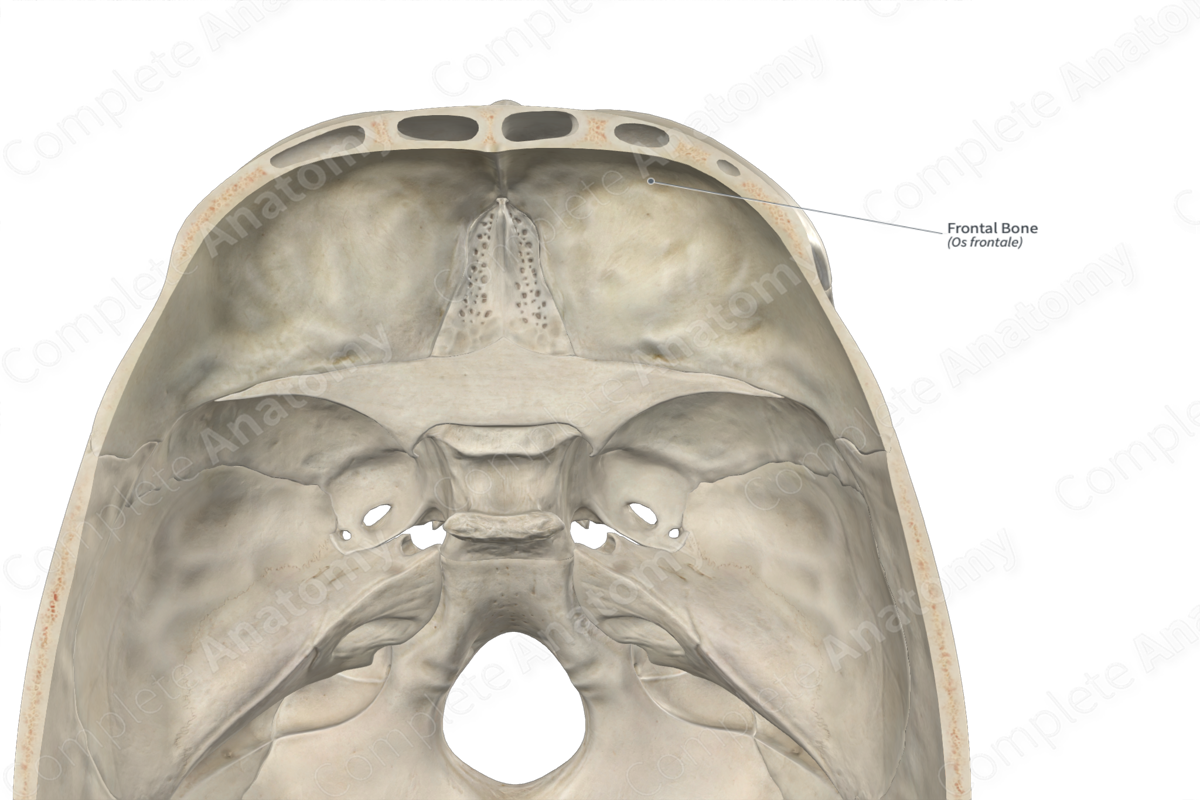
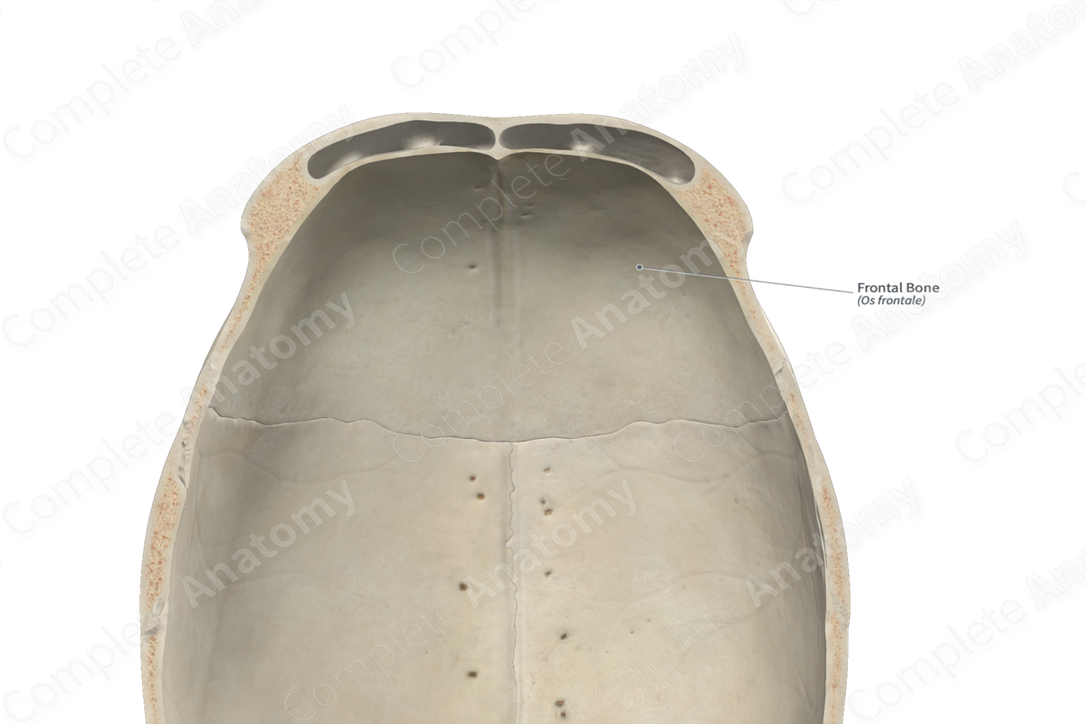
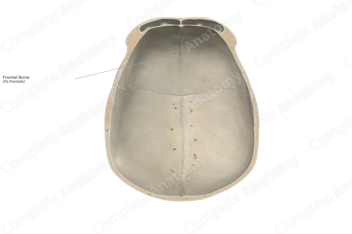
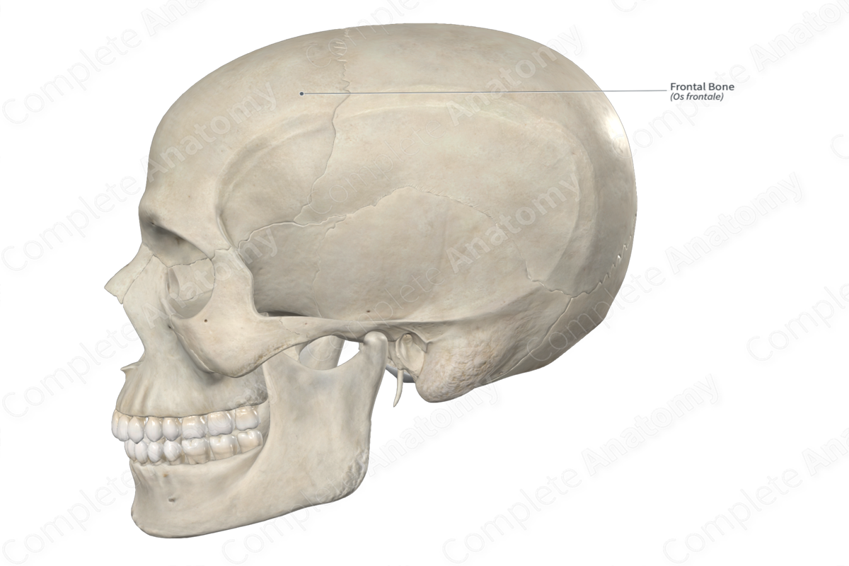
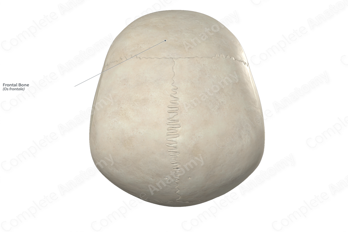
The frontal bone is the single, large bone found along the anterosuperior aspect of the cranium. It is classified as a flat bone, forms the forehead, and contributes to the formation of the neurocranium. The frontal bone consists of right and left frontal sinuses and includes the following bony features:
- parts: squamous, orbital, and nasal parts;
- surfaces: external, internal, orbital, and cerebral surfaces, and parietal, sphenoidal, nasal, and supraorbital margins;
- landmarks: superciliary arches, fossae for lacrimal glands, zygomatic processes, groove for the superior sagittal sinus, frontal eminences, and temporal lines.
More information regarding these bony features can be found in the Parts, Surfaces and Landmarks tabs for this bone.
The frontal bone is located:
- anterior to the parietal bones;
- superior to the sphenoid, ethmoid, zygomatic, lacrimal, and nasal bones, and the maxilla.
It articulates with the:
- parietal bones at the coronal suture;
- ethmoid bone at the frontoethmoidal suture;
- lacrimal bones at the frontolacrimal sutures;
- maxillae at the frontomaxillary sutures;
- nasal bones at the frontonasal suture;
- zygomatic bones at the frontozygomatic sutures;
- sphenoid bone at the sphenofrontal sutures.
Ossification
Ossification of the frontal bone occurs at four ossification centers, these are found in the:
- right and left frontal eminences, with one center found in each, which appear in utero during the second month;
- nasal spine, with two centers found here, which appear during the tenth year.
At birth, the right and left halves of the frontal bone remains unfused, with a metopic suture (frontal suture) located between them. The right and left halves of the frontal bone fuse within the first year after birth(Standring, 2020).
Variations
In some individuals:
- the frontal sinus may be absent unilaterally or bilaterally;
- the metopic suture may still be present in adults (Tubbs, Shoja and Loukas, 2016);
- the frontal and temporal bones meet each other and articulate, resulting in the parietal and sphenoid bones not meeting and articulating.
The frontal bone also displays sexual dimorphism. The male frontal bone has thicker and more rounded orbital margins, more pronounced supraorbital margins, and a usually well-defined glabella. Additionally, the temporal lines are heavier and the supramastoid crest extends posterior to the external acoustic meatus, when compared with the female. The female forehead tends to be higher, more vertical, and more rounded than the male (Standring, 2020; White and Folkens, 2005).
Surface Anatomy
Regarding surface anatomy, the frontal eminences, superciliary arches, supraorbital margins, and temporal lines of the frontal bone can all be palpated.
List of Clinical Correlates
- Fracture of frontal bone
- Hyperostosis frontalis interna
- Craniosynostosis
References
Standring, S. (2020) Gray's Anatomy: The Anatomical Basis of Clinical Practice. 42nd edn.: Elsevier Health Sciences.
Tubbs, R. S., Shoja, M. M. and Loukas, M. (2016) Bergman's Comprehensive Encyclopedia of Human Anatomic Variation. Wiley.
White, T. D. and Folkens, P. A. (2005) The Human Bone Manual. Elsevier Science.
Learn more about this topic from other Elsevier products

