Structure
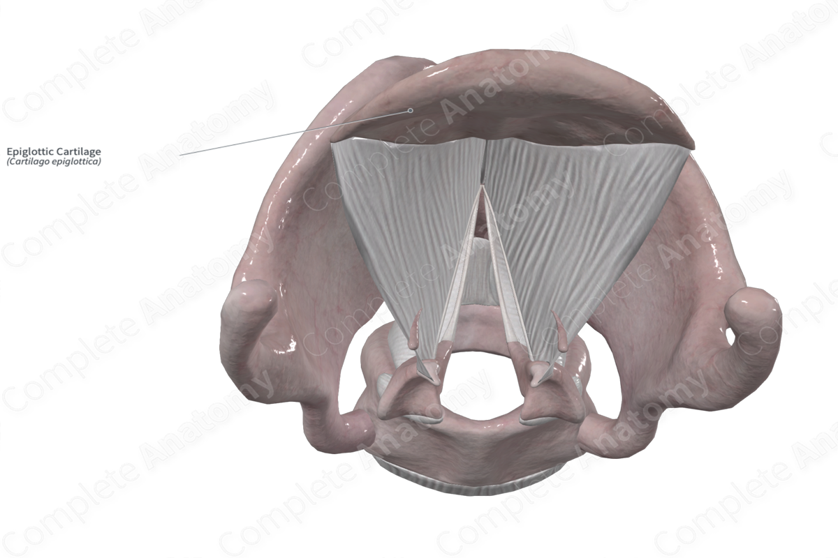
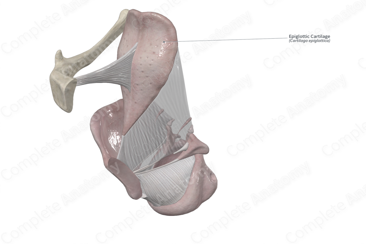
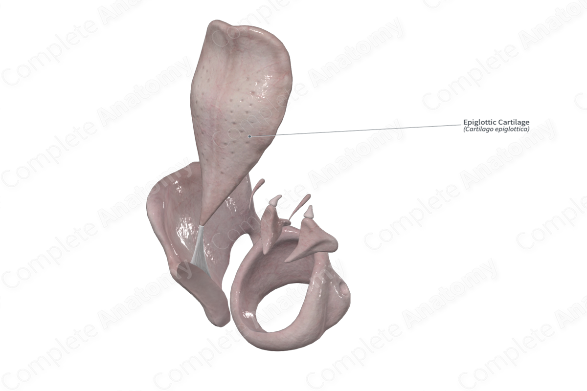
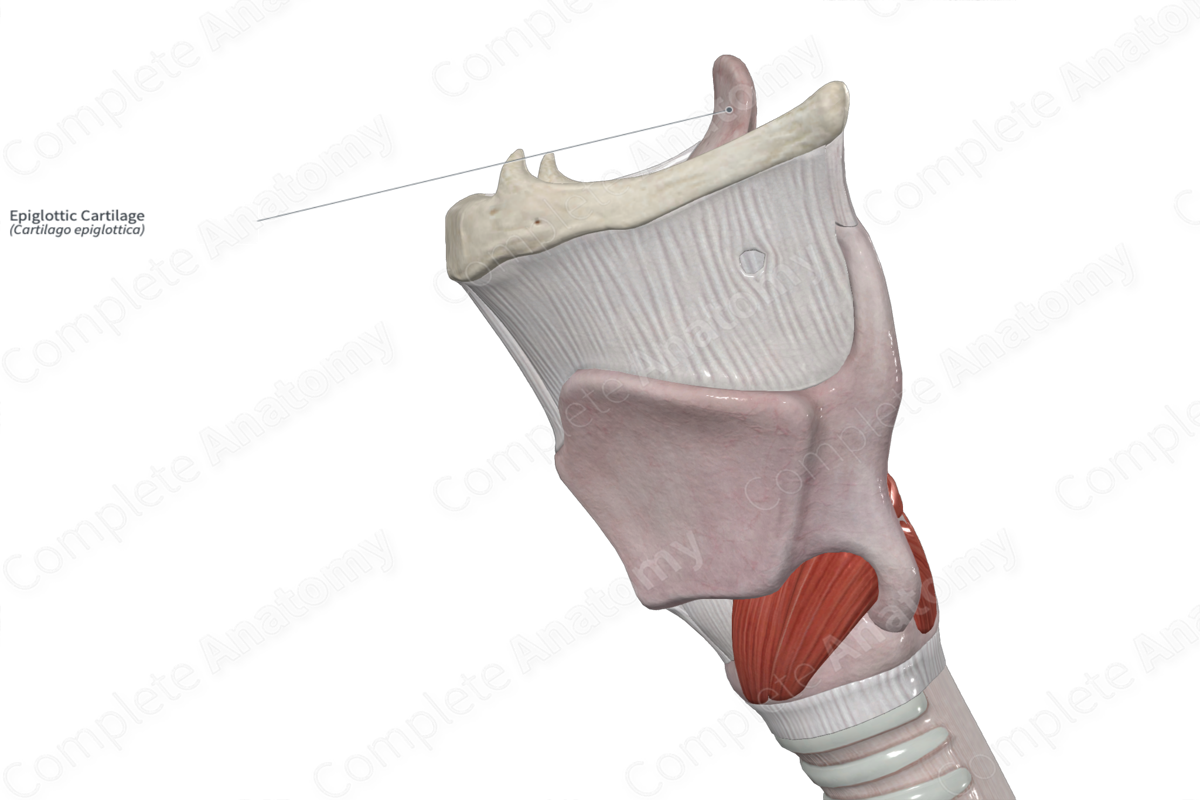
The epiglottis is a flap of elastic cartilage that projects in an oblique fashion posterior to hyoid bone and the tongue. Superiorly, it has a free margin but the inferior portion is attached to the thyroid cartilage via the thyroepiglottic ligament. Laterally, it is connected to the arytenoid cartilages via a mucosal fold called the aryepiglottic fold. Deep to this fold is the aryepiglottic muscle.
The epiglottis has an anterior and a posterior surface.
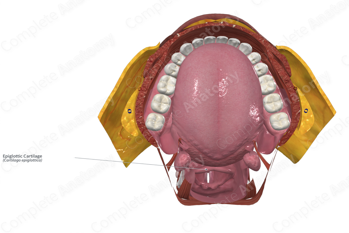
- The inferior portion of the anterior surface is connected to the hyoid bone via a hyoepiglottic ligament. It is also connected to the pharyngeal surface of the tongue and the lateral pharynx by a median and two lateral glossoepiglottic ligaments, respectively. Between the median and lateral glossoepiglottic ligaments are small depressions, the valleculae.
- The posterior surface of the epiglottis is smooth and coated with respiratory mucosa containing numerous small mucus glands. It is pierced by branches of the internal laryngeal nerve.
Function
The closure of the epiglottis ensures food passes towards the lateral food channels. The mechanism behind closure of the airways by the epiglottis remains unclear. Upon deglutition, the hyoid bone positions itself in anterosuperior manner. This results in passive pressure passing through the base of the tongue and “bends” the epiglottis posteriorly. Additionally, aryepiglottic contraction aids the posterior movement of the epiglottis. Furthermore, it has been suggested that the weight of a bolus of food or liquid on the surface of the epiglottis may also aid closure (Standring, 2016).
References
Standring, S. (2016) Gray's Anatomy: The Anatomical Basis of Clinical Practice. Gray's Anatomy Series 41st edition edn.: Elsevier Limited.
Learn more about this topic from other Elsevier products

