Structure
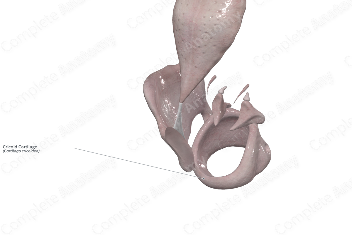
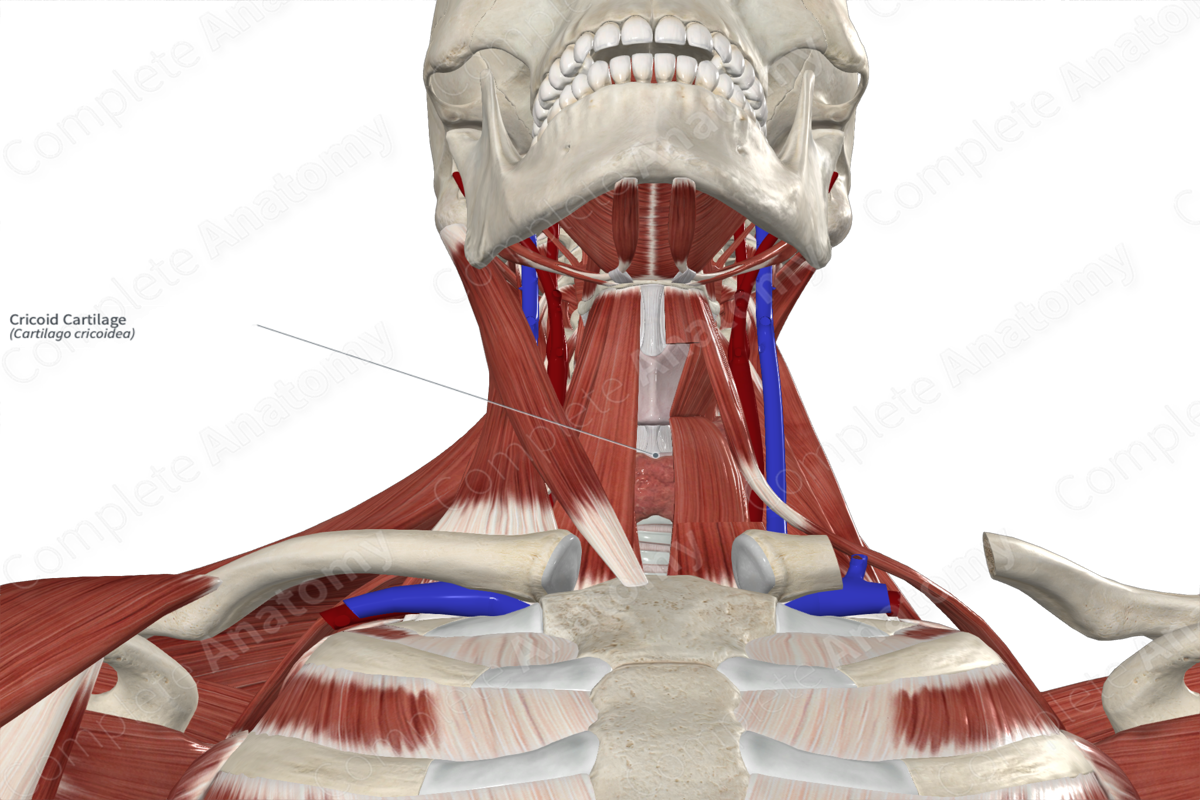
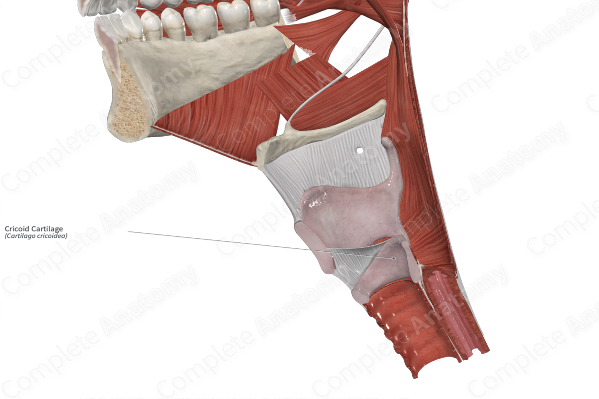
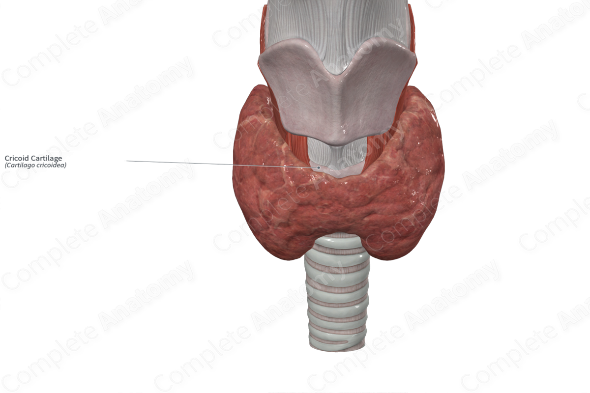
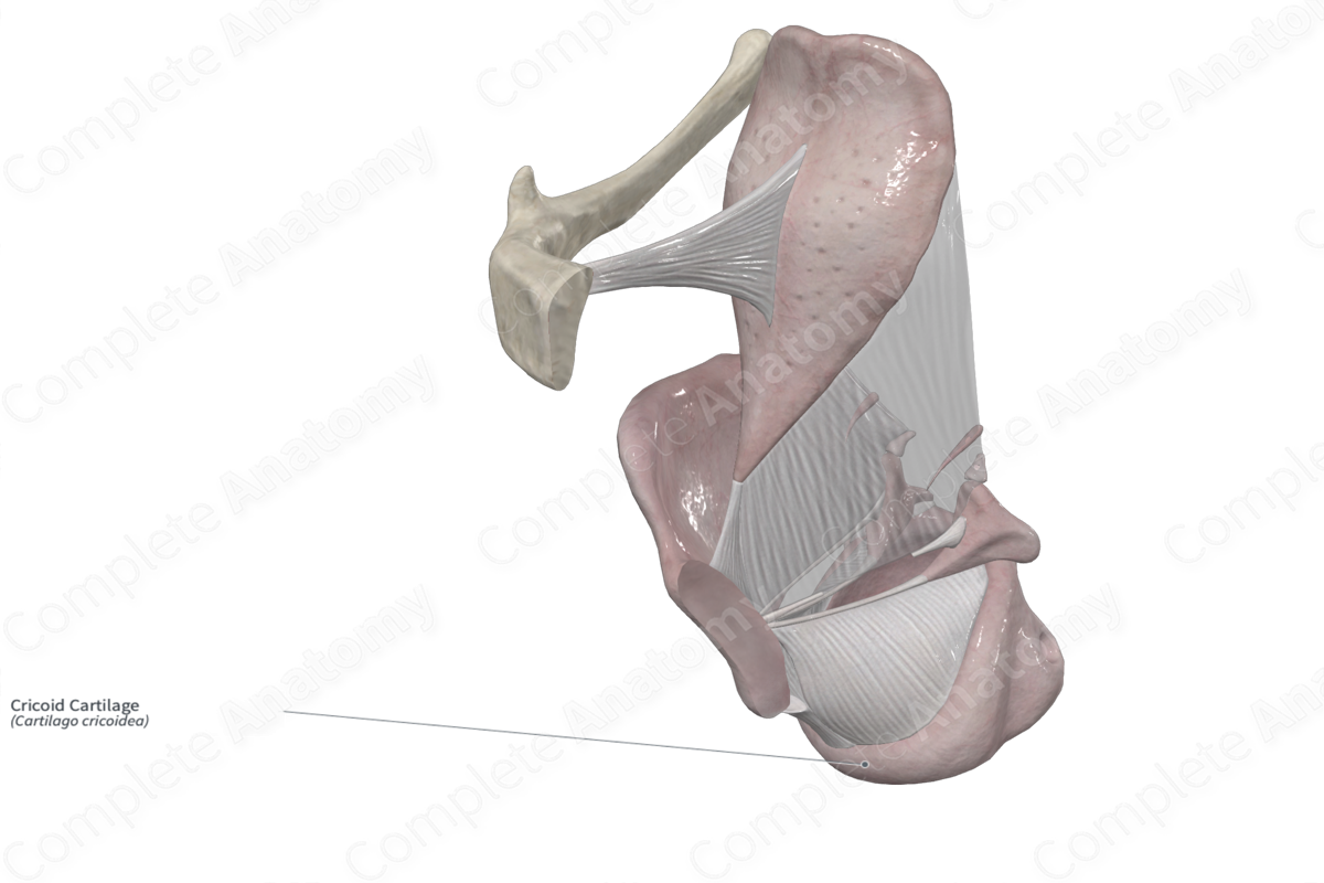
The cricoid cartilage is composed of hyaline cartilage and sits between the thyroid and arytenoid cartilages above and the trachea below. It consists of an anterior cricoid arch and a posterior cricoid lamina and is the only laryngeal cartilage that forms a full circular ring.
Key Features & Anatomical Relations
The cricoid arch is approximately 0.5 cm in height and expands posteriorly as it meets the cricoid lamina. It is the site of attachment for the cricothyroid muscle (an intrinsic laryngeal muscle) and the cricopharyngeal portion of the inferior pharyngeal constrictor muscle.
The cricoid lamina is approximately 2–3 cm in dimension. It has a vertical ridge along the median plane and two concavities on each side. The muscular fibers of the longitudinal layer of the esophagus attach to the upper portion of the ridge. Additionally, some intrinsic laryngeal muscles, such as the cricoarytenoid muscle, attach to the cricoid lamina.
A facet at the junction between the cricoid lamina and the cricoid arch demarcates the location of the cricothyroid joint. Additionally, the superoposterior border has a “shallow” facet for the articulation with the arytenoid cartilage (Standring, 2016).
References
Standring, S. (2016) Gray's Anatomy: The Anatomical Basis of Clinical Practice. Gray's Anatomy Series 41st edition edn.: Elsevier Limited.
Learn more about this topic from other Elsevier products

