Quick Facts
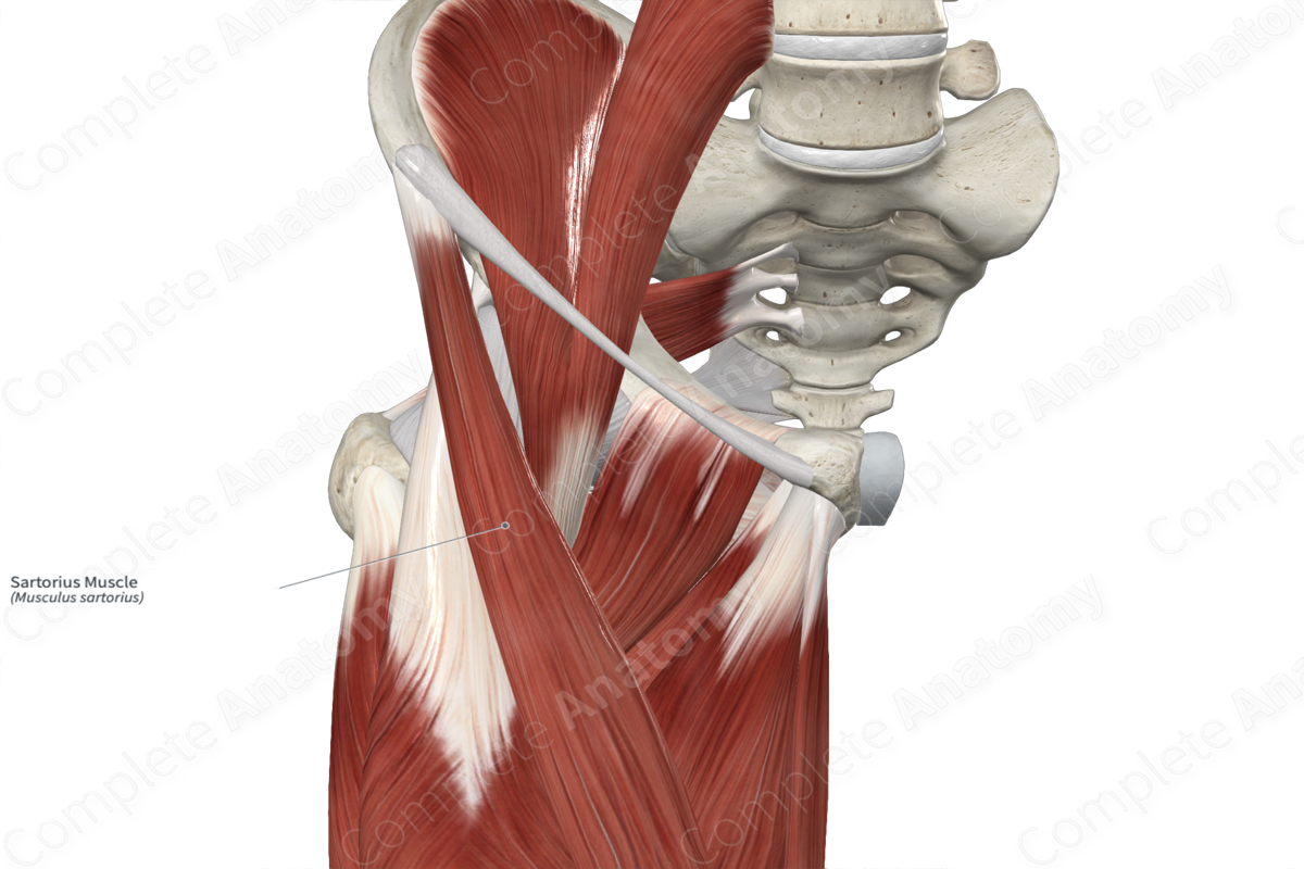
Origin: Anterior superior iliac spine.
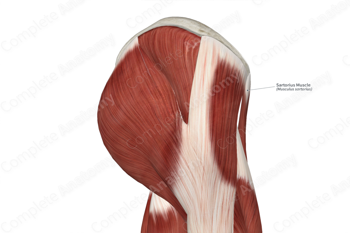
Insertion: Medial aspect of proximal part of tibia.
Action: Flexes and medially rotates leg at knee joint; assists in flexion, abduction and lateral rotation of thigh at hip joint.
Innervation: Femoral nerve (L2-L3).
Arterial Supply: Femoral artery.
Origin
The sartorius muscle originates from the:
- anterior superior iliac spine;
- superior portion of the notch that is located inferior to the anterior superior iliac spine.
Insertion
The fibers of the sartorius muscle travel inferomedially and insert onto the medial aspect of the proximal part of tibia.
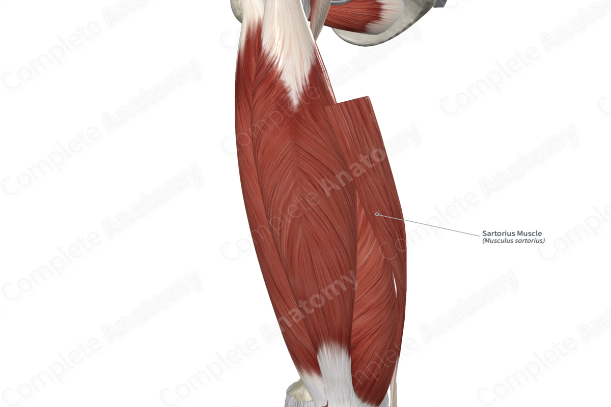
Key Features & Anatomical Relations
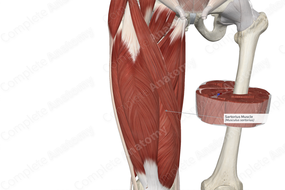
The sartorius muscle is found in the anterior compartment of the thigh. It is the longest muscle in the body and is a narrow, strap-like type of skeletal muscle. In the distal one third of the thigh, the muscle belly gives rise to a narrow, flat tendon, which travels inferiorly. It passes medial to the knee joint and anterior to the tendon of the gracilis muscle, after which it expands into an aponeurosis, which travels to its insertion site. At their insertion sites, the tendons of the sartorius, gracilis and semitendinosus muscles collectively form the pes anserinus, which is Latin for goose’s foot.
The sartorius muscle is located:
- anterior to the rectus femoris, adductor longus, vastus medialis and gracilis muscles;
- medial to the tensor fasciae latae muscle (at its proximal end);
- lateral to the iliopsoas muscle (at its proximal end).
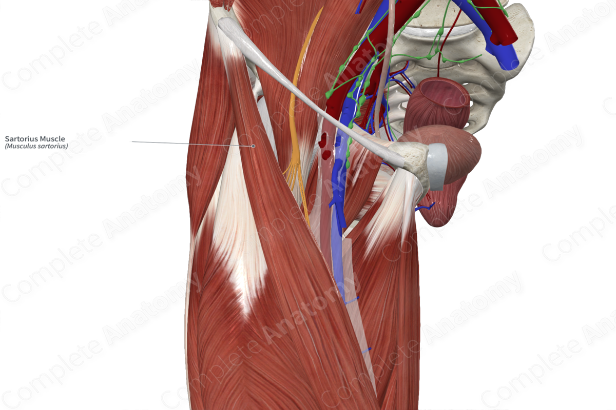
The sartorius muscle contributes to the formation of the:
- adductor canal, where the muscle forms its medial boundary;
- femoral triangle, where the muscle forms its lateral boundary.
Actions & Testing
The sartorius muscle is involved in multiple actions:
- It medially rotates the leg at the knee joint while this joint is held in a flexed position;
- It assists in flexion of the leg at the knee joint;
- It assists in flexion of the thigh at the hip joint;
- It assists in abduction of the thigh at the hip joint;
- It assists in lateral rotation of the thigh at the hip joint (Standring, 2016).
The sartorius muscle can be tested by simultaneously flexing, abducting and laterally rotating the hip against resistance while sitting and the leg is held in a flexed position, during which the muscle can be seen and palpated (Sinnatamby, 2011).
List of Clinical Correlates
- Cruciate ligament surgery
References
Sinnatamby, C. S. (2011) Last's Anatomy: Regional and Applied. ClinicalKey 2012: Churchill Livingstone/Elsevier.
Standring, S. (2016) Gray's Anatomy: The Anatomical Basis of Clinical Practice. Gray's Anatomy Series 41st edn.: Elsevier Limited.
Learn more about this topic from other Elsevier products

