Quick Facts
Origin: Medial part of the intertrochanteric line and medial to the spiral line and linea aspera of the femur, extending inferiorly to the medial supracondylar line.
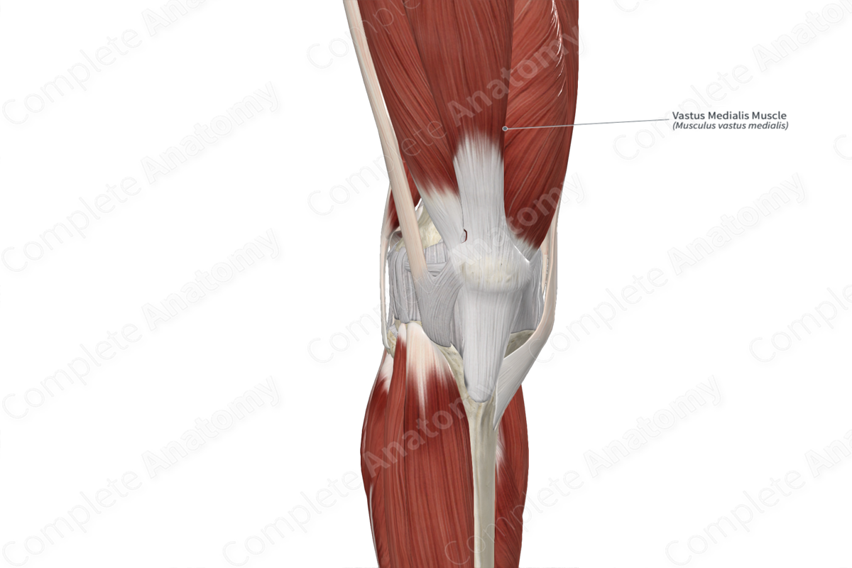
Insertion: Tibial tuberosity, via tendon of quadriceps femoris muscle and patellar ligament, and medial border of patella.
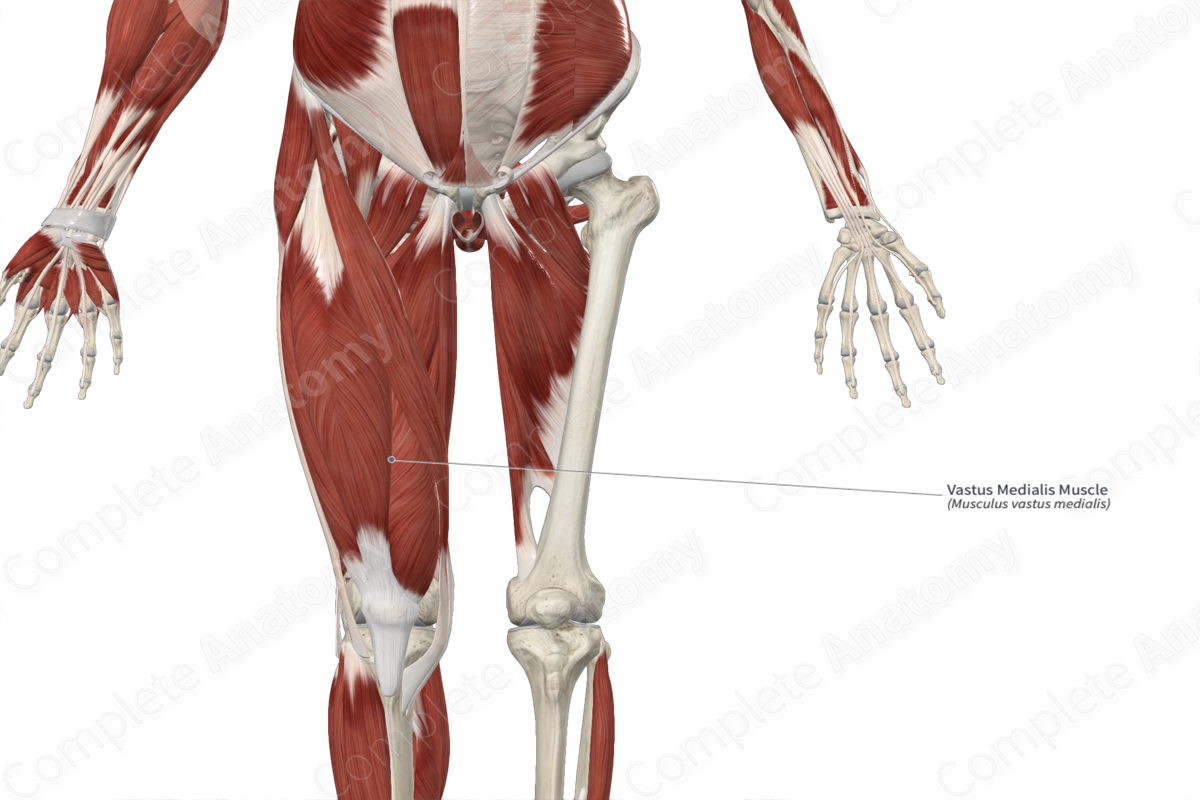
Action: Extends leg at knee joint.
Innervation: Femoral nerve (L2-L4).
Arterial Supply: Femoral artery.
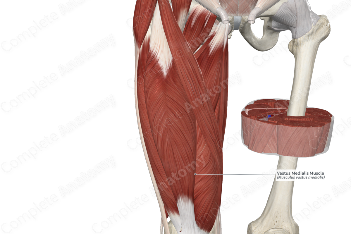
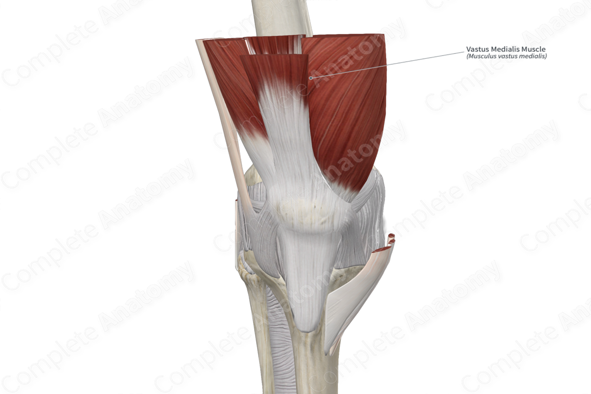
Origin
The vastus medialis muscle originates from the:
- intertrochanteric line of femur;
- medial lip of linea aspera of femur;
- tendon of adductor magnus muscle.
Insertion
The fibers of the vastus medialis muscle travel anteroinferiorly and converge with the fibers of the rectus femoris, vastus intermedius and vastus lateralis muscles to form the tendon of quadriceps femoris muscle. The fibers of this tendon travel superficial to the patella, where they become continuous with the patellar ligament, which inserts onto the tibial tuberosity. Some fibers of the vastus medialis muscle insert directly onto the medial border of the patella.
Key Features & Anatomical Relations
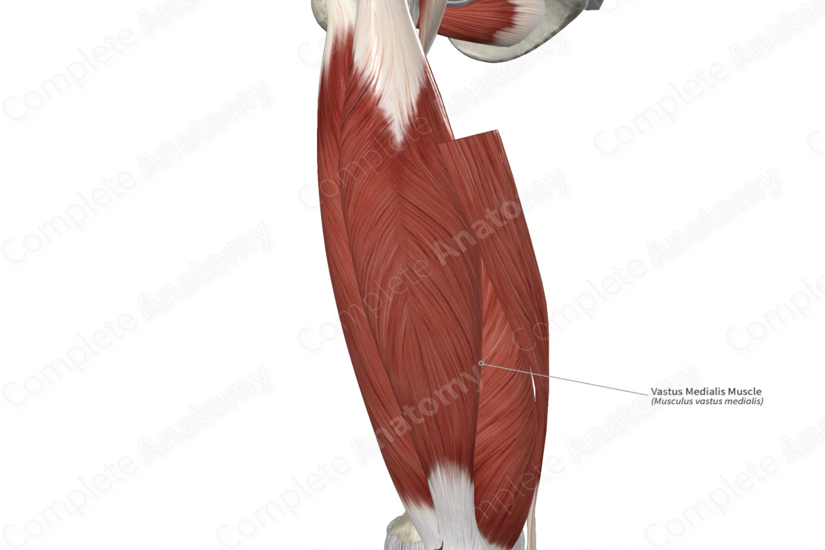
The vastus medialis muscle is one of the four muscles that form the quadriceps femoris muscle, the other three being the rectus femoris, vastus intermedius and vastus lateralis muscles. It is a long, thick, bipennate type of skeletal muscle.
It is located:
- anterior to the adductor longus and adductor magnus muscles;
- posterior to the sartorius muscle;
- medial to the rectus femoris, vastus intermedius and articularis genus muscles.
The vastus medialis muscle contributes to the formation of the adductor canal, where the muscle forms its anterolateral boundary.
Actions & Testing
The vastus medialis muscle is involved in multiple actions:
- extends the leg at the knee joint, via the tendon of quadriceps femoris muscle and patellar ligament;
- helps stabilize the patella, via fibers that insert directly onto it.
The vastus medialis muscle cannot be tested in isolation, therefore all four muscles of the quadriceps femoris are tested simultaneously by extending the leg at the knee joint against resistance while lying in the supine position with the hip flexed, during which the vastus medialis muscle can be palpated (Standring, 2016).
List of Clinical Correlates
- Patellar instability
- Patellar pain
- Patellar tracking on the femur
References
Standring, S. (2016) Gray's Anatomy: The Anatomical Basis of Clinical Practice. Gray's Anatomy Series 41st edn.: Elsevier Limited.
Learn more about this topic from other Elsevier products

