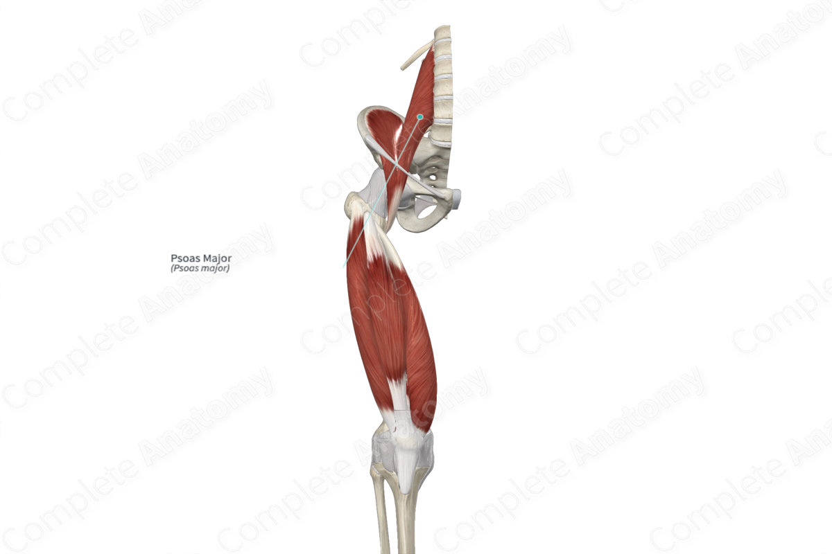
Quick Facts
Origin: Transverse processes of L1-L5 vertebrae, vertebral bodies of T12-L5 vertebrae, and adjacent intervertebral discs.
Insertion: Lesser trochanter of femur.
Action: Flexes thigh at hip joint; flexes trunk.
Innervation: Anterior rami of first, second, and third lumbar nerves.
Arterial Supply: Lumbar arteries, lumbar branch of iliolumbar artery, obturator, external iliac, and femoral arteries.


Related parts of the anatomy
Origin
The psoas major muscle originates from the:
- anteroinferior aspects of the transverse processes of the lumbar vertebrae;
- anterolateral aspects of the vertebral bodies of the twelfth thoracic to fifth lumbar vertebrae;
- adjacent intervertebral discs.
There can be variations between individuals regarding the numbers of origin sites and the vertebrae to which they attach.
Insertion
The fibers of the psoas major muscle travel anteroinferiorly and insert, via a thick, rounded tendon, onto the lesser trochanter of the femur.
Key Features & Anatomical Relations
The psoas major muscle is one of the two muscles that form the iliopsoas muscle, the other being the iliacus muscle. It is a long, thick, fusiform type of skeletal muscle. Within the posterior abdominal wall, the fibers of the psoas major muscle converge as they travel anteroinferiorly towards the inguinal ligament. After it passes deep to the middle one third of the inguinal ligament, the psoas major muscle travels inferiorly within the anterior compartment of the thigh. Within the proximal region of this compartment, the muscle belly gives rise to a thick, rounded tendon, which travels posteroinferiorly to its insertion site. Furthermore, the tendon of the psoas major provides an insertion site for the iliacus muscle.
The psoas major muscle is located:
- anterior to the capsule of the hip joint, the quadratus lumborum muscle, and the iliopectineal bursa;
- posterior to the ureter and inguinal ligament;
- medial to the kidney, the iliacus muscle, and the femoral nerve;
- lateral to the lumbar vertebrae and the psoas minor and pectineus muscles.
The lumbar plexus lies within the posterior portion of the psoas major muscle, with its branches emerging from the borders and surfaces of the muscle.
Actions & Testing
Overall, the iliopsoas muscle is involved in multiple actions:
- during unilateral contraction, it flexes the thigh at the hip joint;
- during bilateral contraction, it flexes the trunk.
The psoas major muscle cannot be tested in isolation, therefore both muscles of the iliopsoas are tested simultaneously by flexing the thigh at the hip joint against resistance while in the supine position with the knee flexed (Standring, 2016).
List of Clinical Correlates
- Iliopsoas tendinitis
- Thomas test
- Psoas sign
References
Standring, S. (2016) Gray's Anatomy: The Anatomical Basis of Clinical Practice. Gray's Anatomy Series 41st edn.: Elsevier Limited.
Learn more about this topic from other Elsevier products

