Quick Facts
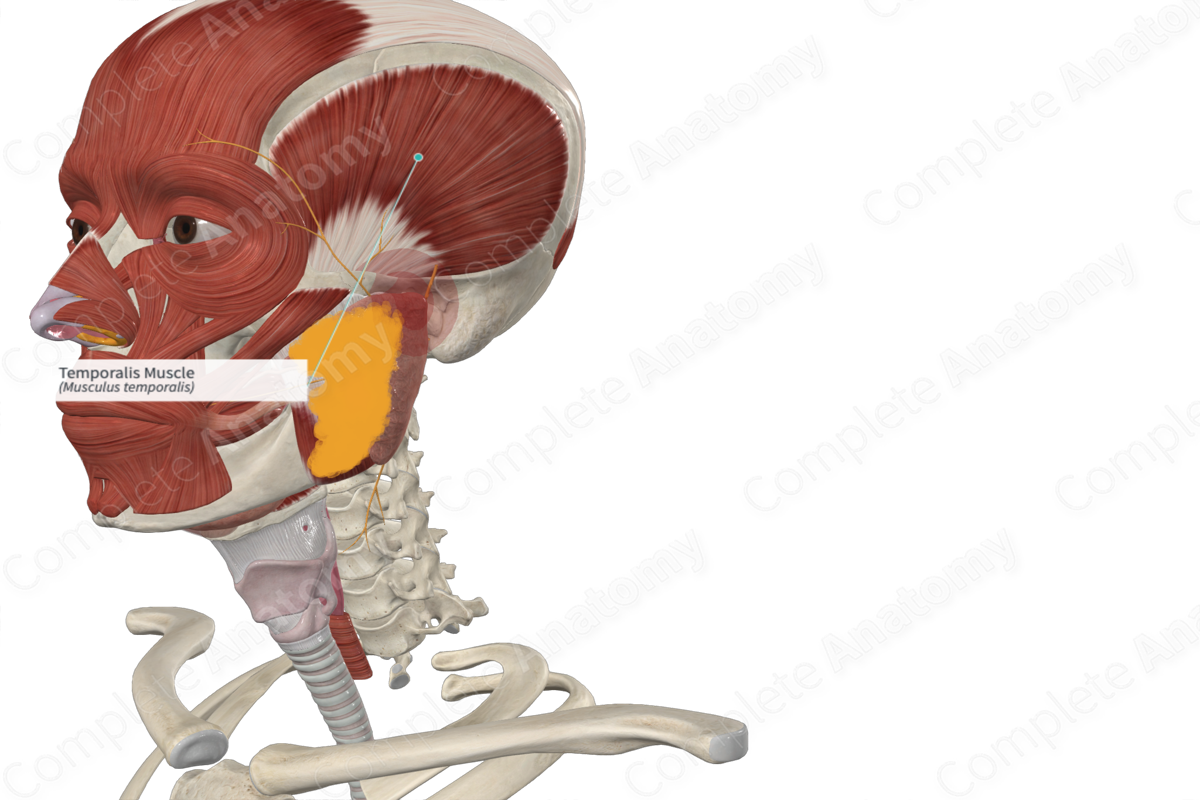
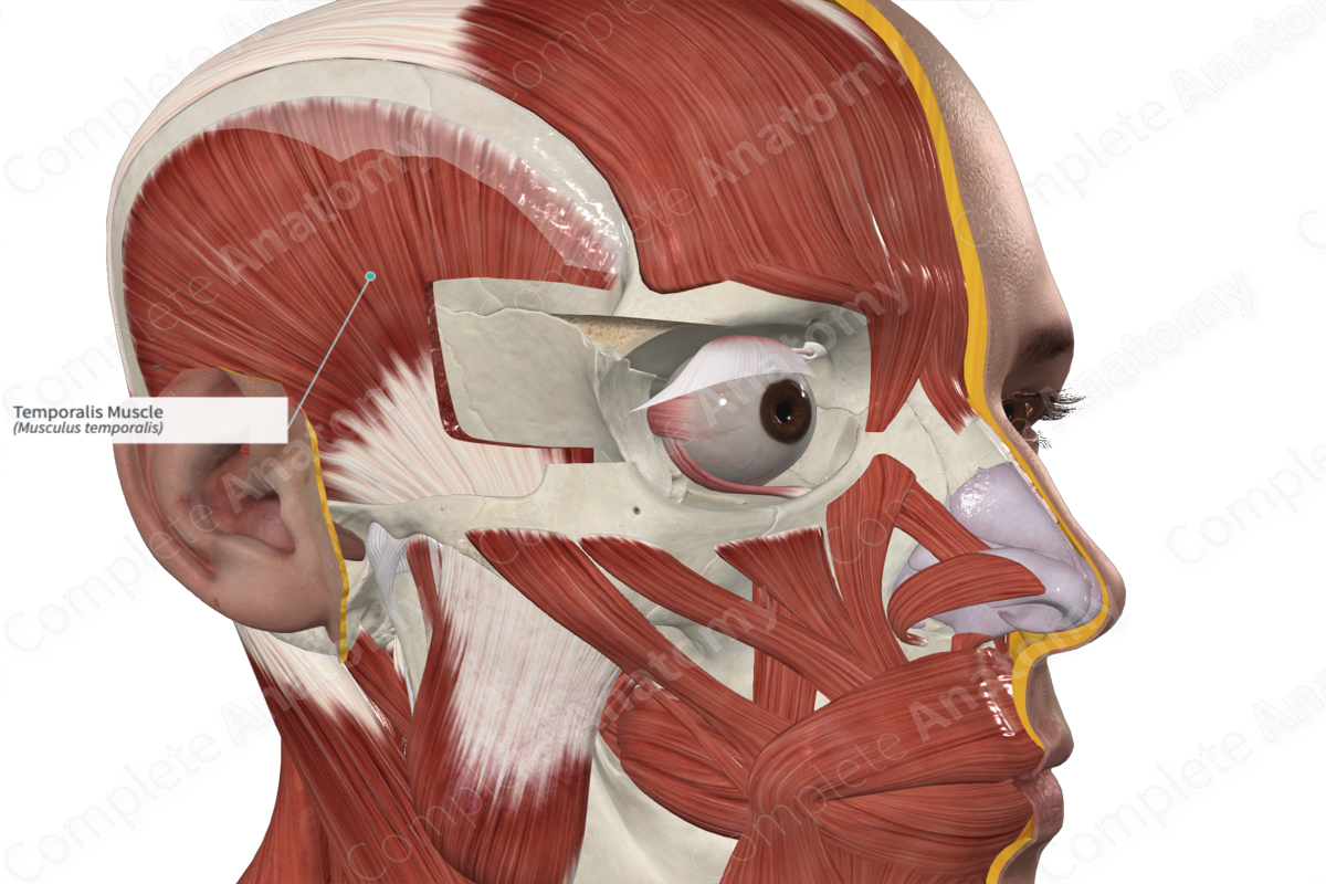
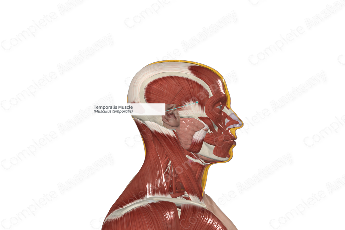
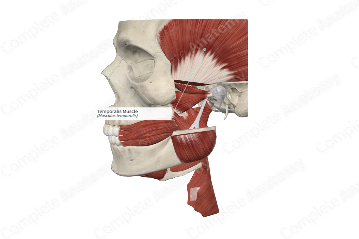
Origin: Temporal fossa and fascia.
Insertion: Coronoid process and ramus of mandible.
Action: Elevates and retracts mandible.
Innervation: Anterior and posterior deep temporal nerves (CN V3).
Arterial Supply: Anterior and posterior deep temporal arteries and middle temporal artery.
Origin
The temporalis muscle originates from the temporal fossa and its associated temporal fascia.
Insertion
The fibers of the temporalis muscle converge as they descend deep to the zygomatic arch and attach to the coronoid process and ramus of the mandible.
Key Features & Anatomical Relations
The temporal fossa sits deep to the temporalis muscle. Within this fossa are the deep temporal arteries, veins, and nerves.
The fibers of the anterior portion of the temporalis muscle run in an approximately vertical direction, which permits elevation of the mandible. In contrast, the posterior component has fibers which run almost horizontally. This permits retraction of the mandible.
Actions
The temporalis muscle is involved in multiple actions:
- elevates the mandible at the temporomandibular joint;
- retracts the mandible at the temporomandibular joint (Standring, 2016).
List of Clinical Correlates
- Trismus
References
Standring, S. (2016) Gray's Anatomy: The Anatomical Basis of Clinical Practice. Gray's Anatomy Series 41st edn.: Elsevier Limited.
Learn more about this topic from other Elsevier products

