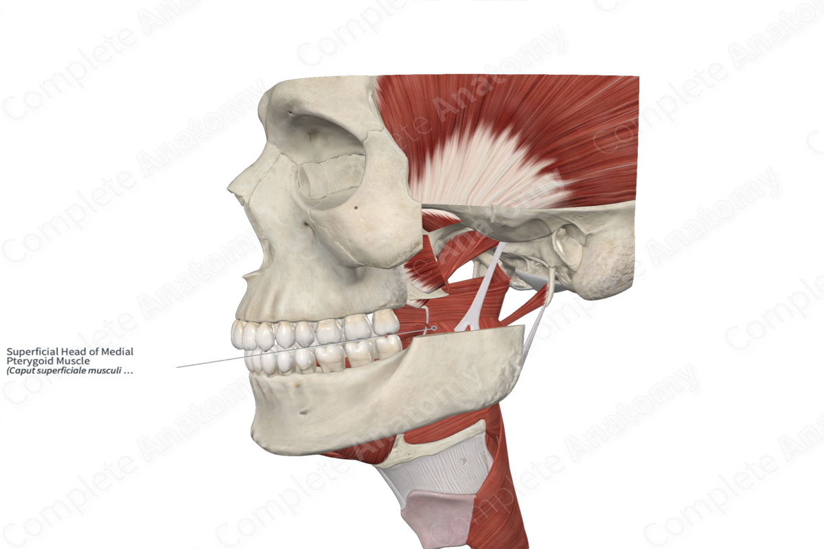
Superficial Head of Medial Pterygoid Muscle
Caput superficiale musculi pterygoidei medialis
Read moreQuick Facts
Origin: Maxillary tuberosity; pyramidal process of palatine bone.
Insertion: Medial surface of ramus and angle of mandible.
Action: Laterally moves and protracts mandible.
Innervation: Nerve to medial pterygoid muscle (CN V3).
Arterial Supply: Pterygoid branches of maxillary artery.


Related parts of the anatomy
Origin
The medial pterygoid muscle is a quadrangular muscle that has two heads. The smaller superficial head of the medial pterygoid muscle originates from the maxillary tuberosity and the pyramidal process of the palatine bone.
Insertion
The fibers of the medial pterygoid course in a posterolateral direction and attach to the medial surface of the angle and ramus of the mandible via a strong tendinous lamina.
Key Features & Anatomical Relations
The medial pterygoid muscle mainly elevates the jaw. Since the fibers of the medial pterygoid pass posterolaterally, it may assist the lateral pterygoid muscle in protruding the jaw.
Actions
Overall, the medial pterygoid muscle is involved in multiple actions:
- during unilateral contraction, it laterally moves the mandible to the opposite side at the temporomandibular joint;
- during bilateral contraction, it protracts the mandible at the temporomandibular joint;
- during bilateral contraction, it assists in elevation of the mandible at the temporomandibular joint (Standring, 2016).
List of Clinical Correlates
- Trismus
References
Standring, S. (2016) Gray's Anatomy: The Anatomical Basis of Clinical Practice. Gray's Anatomy Series 41st edn.: Elsevier Limited.
Learn more about this topic from other Elsevier products

