
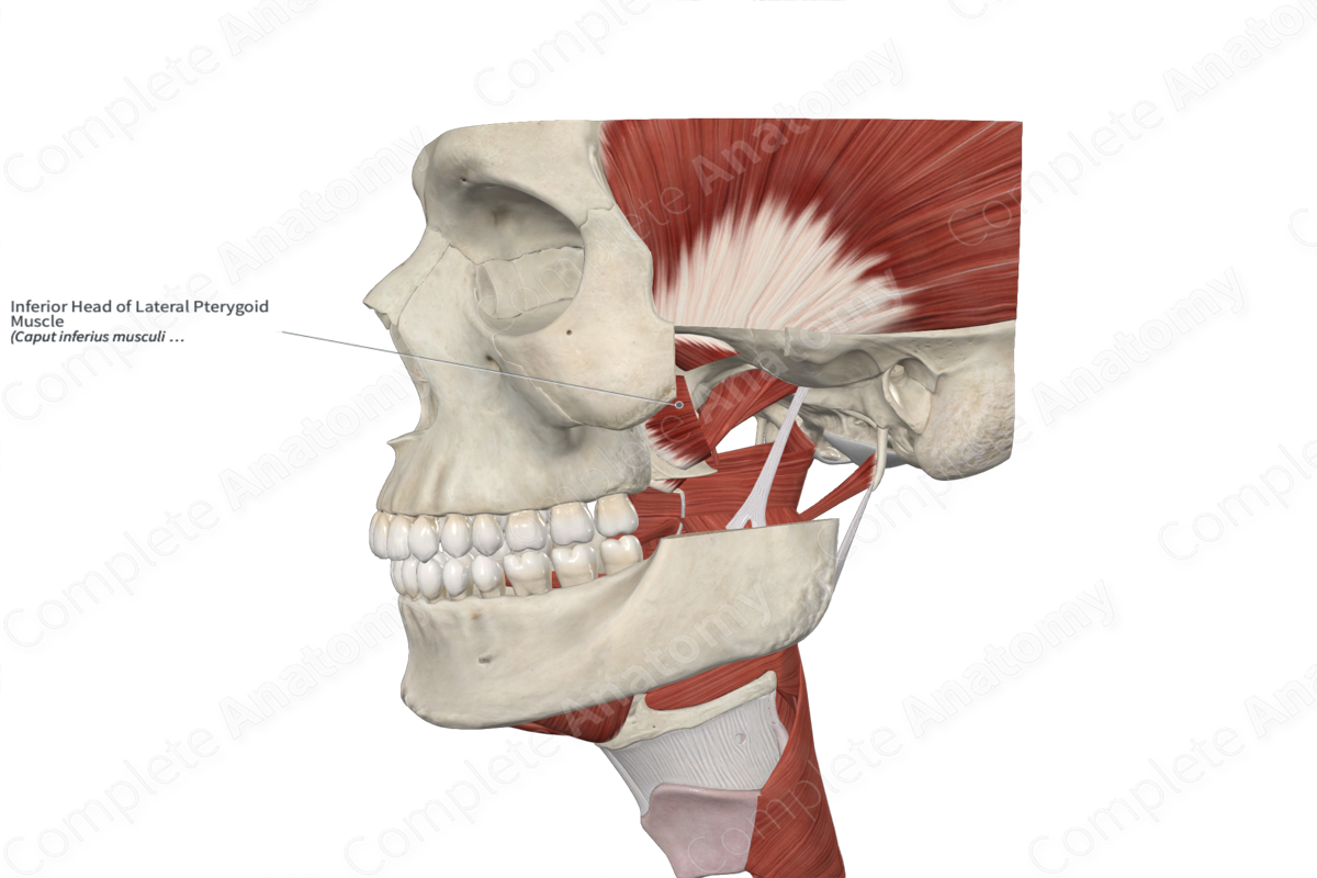
Inferior Head of Lateral Pterygoid Muscle
Caput inferius musculi pterygoidei lateralis
Read moreQuick Facts
Origin: Lateral aspect of lateral pterygoid plate of sphenoid bone.
Insertion: Neck of mandible.
Action: Laterally moves and protracts mandible.
Innervation: Nerve to lateral pterygoid muscle (CN V3).
Arterial Supply: Pterygoid branches of maxillary artery and ascending palatine artery.


Related parts of the anatomy
Origin
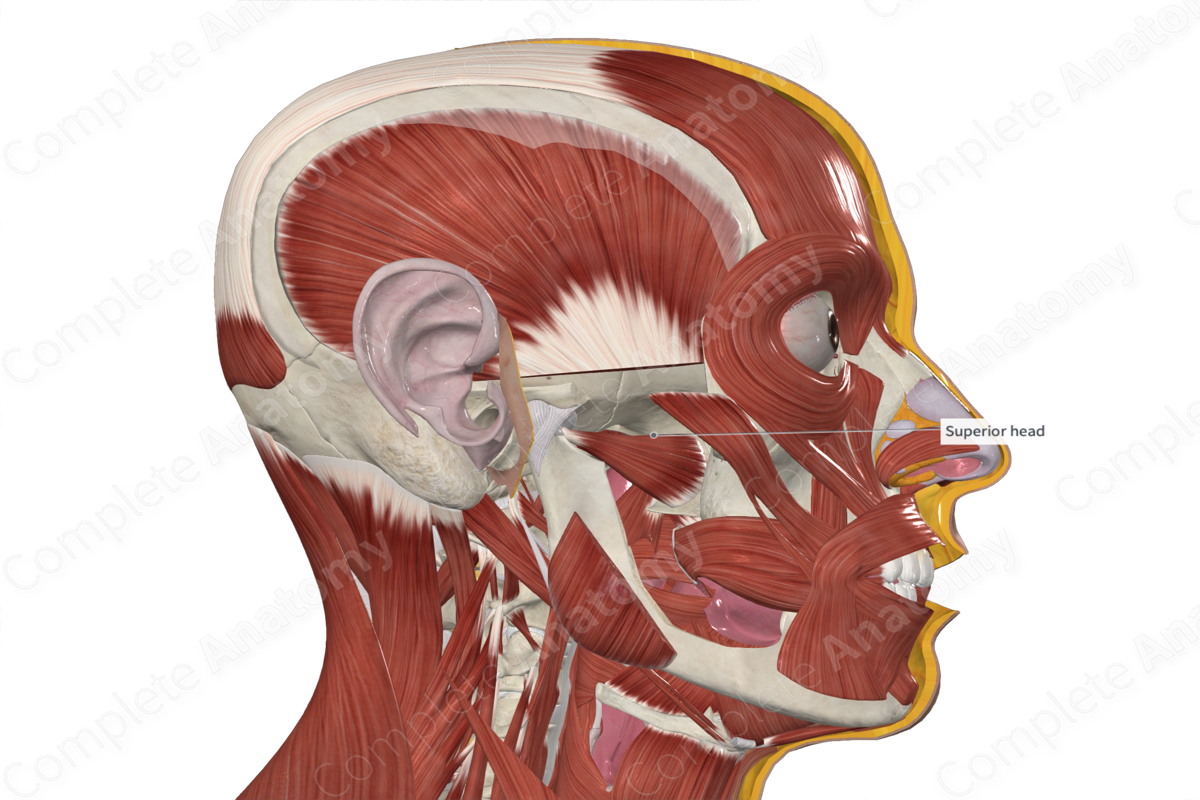
The lateral pterygoid muscle arises as two heads. The inferior head originates from the lateral aspect of the lateral pterygoid plate of the sphenoid bone.
Insertion
The muscular fibers of the lateral pterygoid muscle converge and course in a posterolateral direction to insert into the pterygoid fovea on the neck of the mandibular. Some fibers from the upper head may also attach to the articular disc of the temporomandibular joint (Standring, 2016).
Key Features & Anatomical Relations
The fibers of the lateral pterygoid are orientated horizontally.
Actions
Overall, the lateral pterygoid muscle is involved in multiple actions:
- during unilateral contraction, it laterally moves the mandible to the opposite side at the temporomandibular joint;
- during bilateral contraction, it protracts the mandible at the temporomandibular joint (Standring, 2016).
References
Standring, S. (2016) Gray's Anatomy: The Anatomical Basis of Clinical Practice. Gray's Anatomy Series 41st edn.: Elsevier Limited.
Learn more about this topic from other Elsevier products

