Quick Facts
Origin: Spinous processes of T7-T12 vertebrae, spinous processes of L1-L5 vertebrae via thoracolumbar fascia, ninth to twelfth ribs, iliac crest.
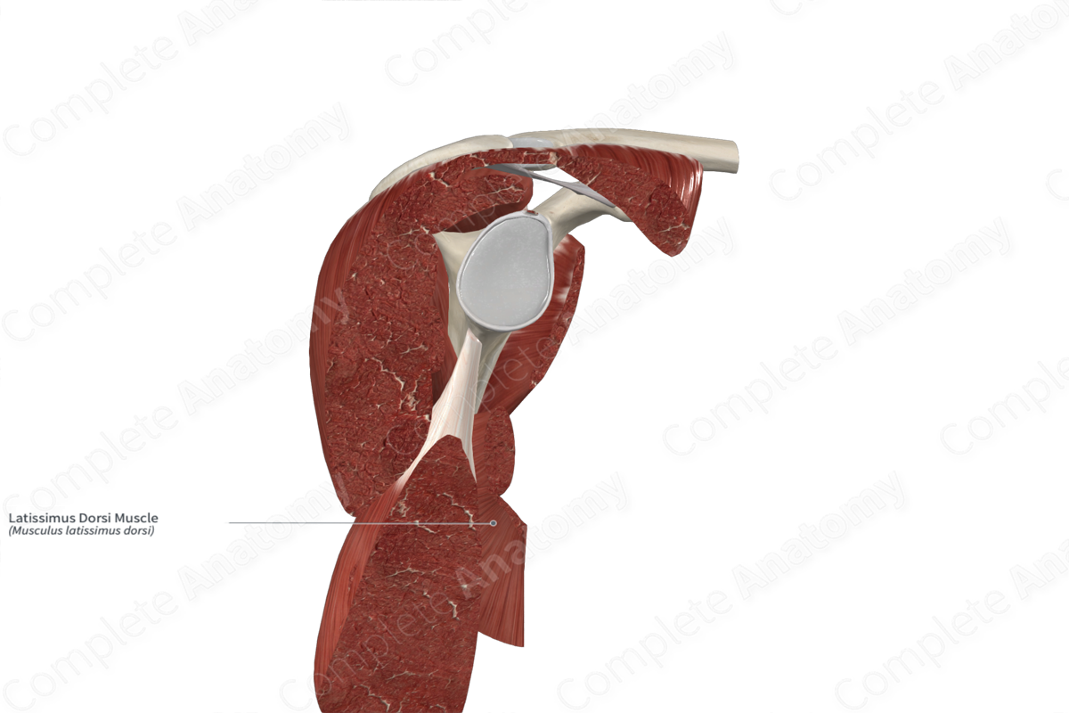
Insertion: Floor of intertubercular sulcus of humerus.
Action: Adducts, medially rotates, and extends arm at glenohumeral (shoulder) joint.
Innervation: Thoracodorsal nerve (C6-C8).
Arterial Supply: Thoracodorsal artery, dorsal branches of ninth to eleventh posterior intercostal, subcostal and first to third lumbar arteries.
Origin
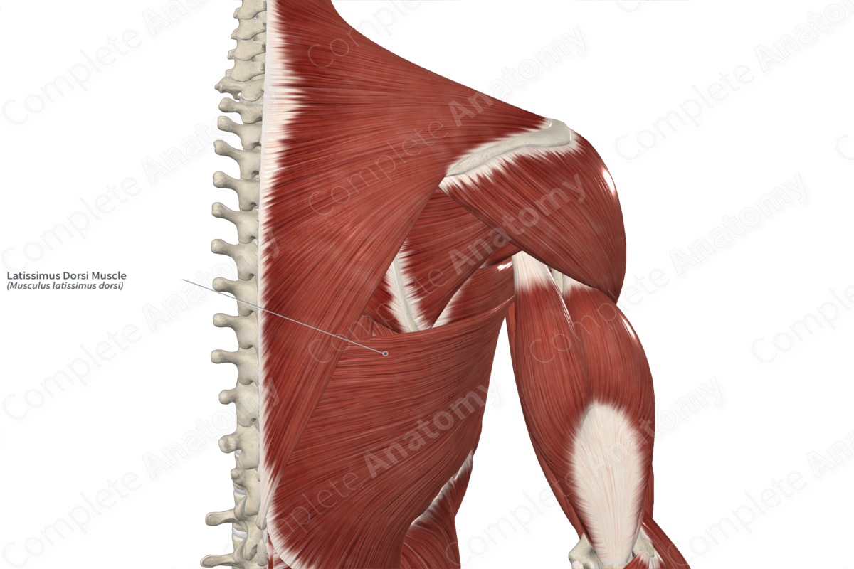
The latissimus dorsi muscle originates from the:
spinous processes of the seventh to twelfth thoracic vertebrae;
spinous processes of the first to fifth lumbar vertebrae, via the posterior layer of the thoracolumbar fascia;
external surfaces of the ninth to twelfth ribs;
posterior half of the iliac crest.
In some individuals, the latissimus dorsi muscle can also originate from the inferior angle of the scapula.
Insertion
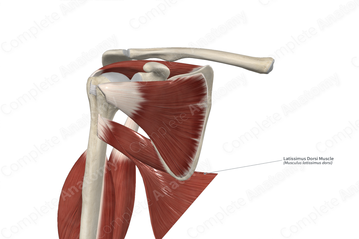
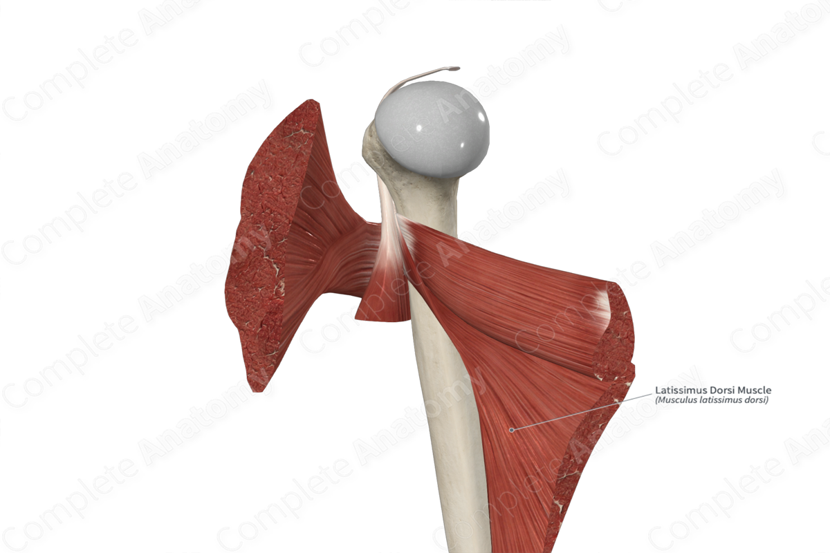
The fibers of the latissimus dorsi muscle travel superolaterally and anteriorly around the thoracic cage. They converge to a single narrow, flat tendon, which inserts onto the floor of the intertubercular sulcus of the humerus.
Proximal to its insertion site, the muscle fibers of the latissimus dorsi spiral. This results in the muscle fibers originating from the most superior location on the back inserting onto the most inferior location on the floor of the intertubercular sulcus of the humerus, and vice versa.
The insertion site for the latissimus dorsi muscle is located between the insertion sites for the teres major and pectoralis major muscles.
Key Features & Anatomical Relations
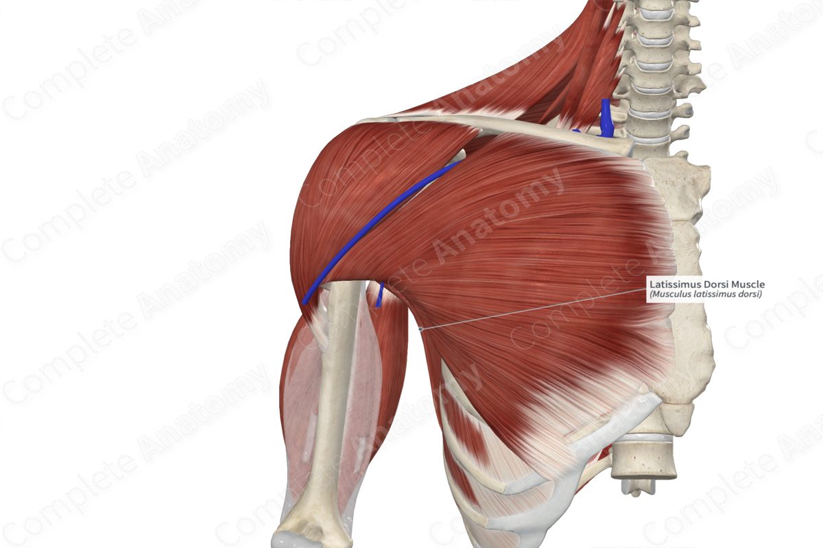
The latissimus dorsi muscle is an extrinsic muscle of the back and is found in the lower back region. It is a broad, flat, convergent type of skeletal muscle.
It is located:
superficial to the inferior angle of scapula, the eighth to twelfth ribs and their adjacent external intercostal muscles, and the serratus anterior, serratus posterior inferior, internal abdominal oblique and erector spinae muscles;
deep to the ascending part of trapezius muscle;
inferior to the teres major, rhomboid major and supraspinatus muscles.
The latissimus dorsi muscle contributes to the formation of the:
posterior wall of the axilla, and its inferior margin contributes to the formation of the posterior axillary fold;
triangle of auscultation, where the latissimus dorsi muscle forms its inferior boundary;
lumbar triangle (triangle of Petit), where the latissimus dorsi muscle forms its posterior boundary.
At their origin sites on the ribs, the fibers of the latissimus dorsi muscle interdigitate with the fibers of the external abdominal oblique muscle.
Actions & Testing
The latissimus dorsi muscle is involved in multiple actions:
adducts the arm at the glenohumeral (shoulder) joint;
medially rotates the arm at the glenohumeral joint;
extends the arm at the glenohumeral joint;
can act as an accessory muscle of respiration.
The latissimus dorsi muscle can be tested by adducting the arm at the glenohumeral joint against resistance, during which the muscle can be seen and palpated (Standring, 2016).
List of Clinical Correlates
Contributes to the formation of triangle of auscultation;
Contributes to the formation of the lumbar triangle, which is a potential site for lumbar herniation.
References
Standring, S. (2016) Gray's Anatomy: The Anatomical Basis of Clinical Practice. Gray's Anatomy Series 41st edn.: Elsevier Limited.
Learn more about this topic from other Elsevier products

