Structure
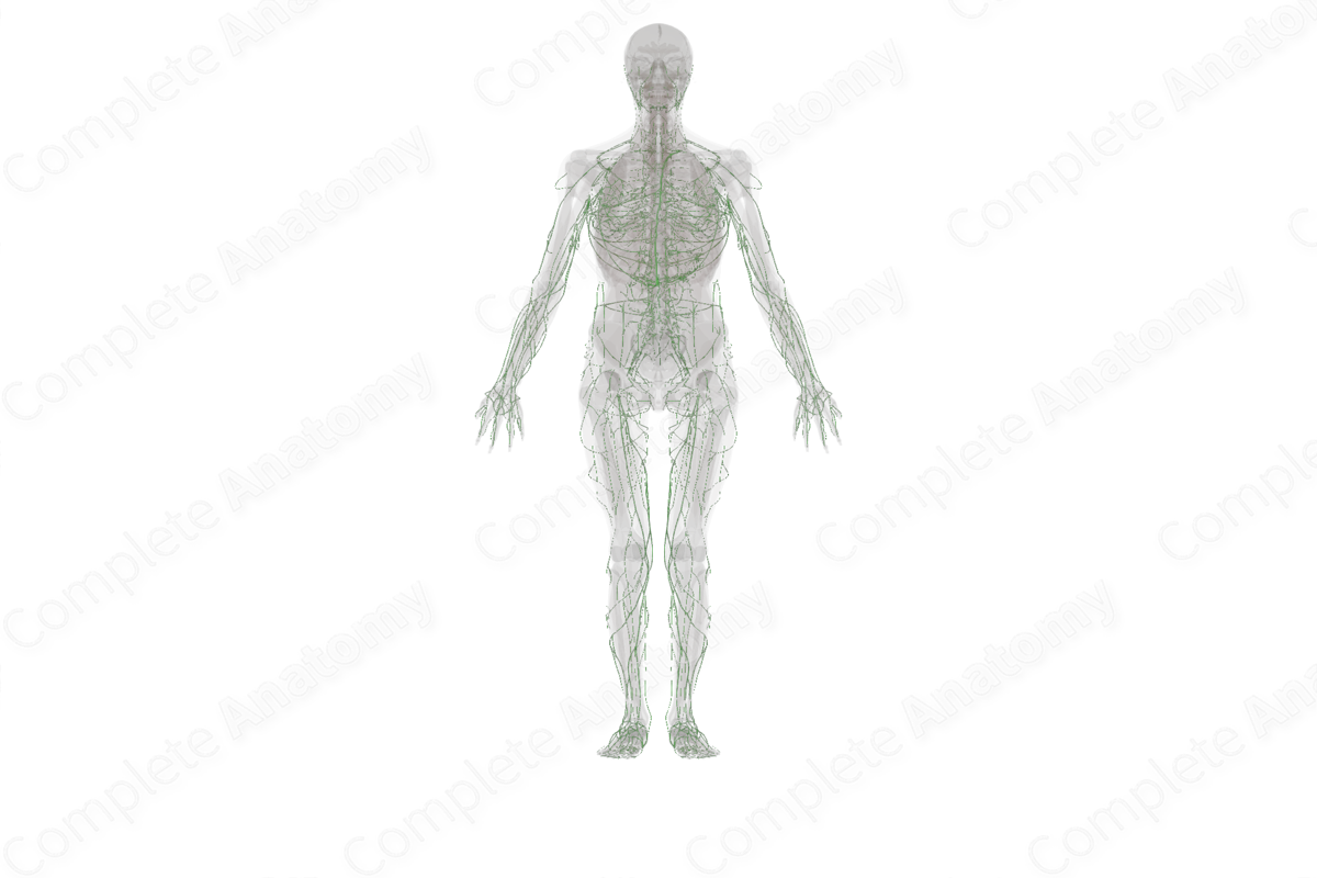
The lymphatic vessels form a secondary circulation system in the human body, the lymphatic circulation. The vessels form a drainage system, usually running parallel with veins.
Afferent lymphatic vessels are formed by the coalescence of blind ended lymphatic capillaries that permeate the vast majority of tissues within the body. Lymphatic capillaries are small with a diameter of about 10–60 µm and comprise a monolayer of endothelial cells that lacks a continuous basal lamina (Margaris and Black, 2012). The unique arrangement of the endothelial cells in capillaries allows them to act as one-way valves, allowing the entry of fluid and substances (such as antigens and cells) from the extracellular spaces of connective tissues, thus producing lymph, and relaying it back to the afferent lymphatic vessels. Each lobule of the lymph node has its own associated afferent vessel. The afferent vessels open through the capsule of the lymph node and deliver lymph to the subcapsular sinus. Since each afferent vessel brings lymph from a different region of tissue and delivers it to its own lobule in the node, it is possible for each lobule to be exposed to different sets of immunological agents, such as antigens, inflammatory mediators, etc. For this reason, cortical and medullary compartments of each lobule may not be uniform in appearance (Willard-Mack, 2006). Afferent lymphatic vessels convey lymph, lymphocytes and antigen-presenting accessory or dendritic cells into the subcapsular, trabecular and medullary sinuses of lymph nodes.
Efferent lymphatic vessels are composed of an internal monolayer of endothelium, surrounded by several layers of smooth muscle, supported by a continuous basal lamina. These lymphatic vessels convey lymph from the lymph node to larger lymph vessels and ducts. The large ducts drain lymph directly into the venous system.
Both afferent and efferent lymphatic vessels also play an important role in homeostasis of extracellular fluid as they drain tissue or interstitial fluid. On average, 8–12 liters of afferent lymph is produced daily, of which 4–8 liters is reabsorbed into the blood in the lymph node. The remaining four liters are transported in efferent lymphatic vessels back to the venous system directly. Accumulation of fluid results in edema of the affected area (Rovenska and Rovensky, 2011).
Each efferent vessel contains valves, which prevents the retrograde or backflow of lymph within the vessel. The valves are bi-leaflet and are composed of a thin layer of collagen between two layers of endothelial cells.


Related parts of the anatomy
References
Margaris, K. N. and Black, R. A. (2012) 'Modelling the lymphatic system: challenges and opportunities', J R Soc Interface, 9(69), pp. 601-12.
Rovenska, E. and Rovensky, J. (2011) 'Lymphatic vessels: structure and function', The Israel Medical Association Journal, 11, pp. 762-768.
Willard-Mack, C. L. (2006) 'Normal structure, function, and histology of lymph nodes', Toxicologic Pathology, 5(34), pp. 409-424.
Learn more about this topic from other Elsevier products
Treatment of the lymphatics: Video, Causes, & Meaning

Treatment of the lymphatics: Symptoms, Causes, Videos & Quizzes | Learn Fast for Better Retention!
