Quick Facts
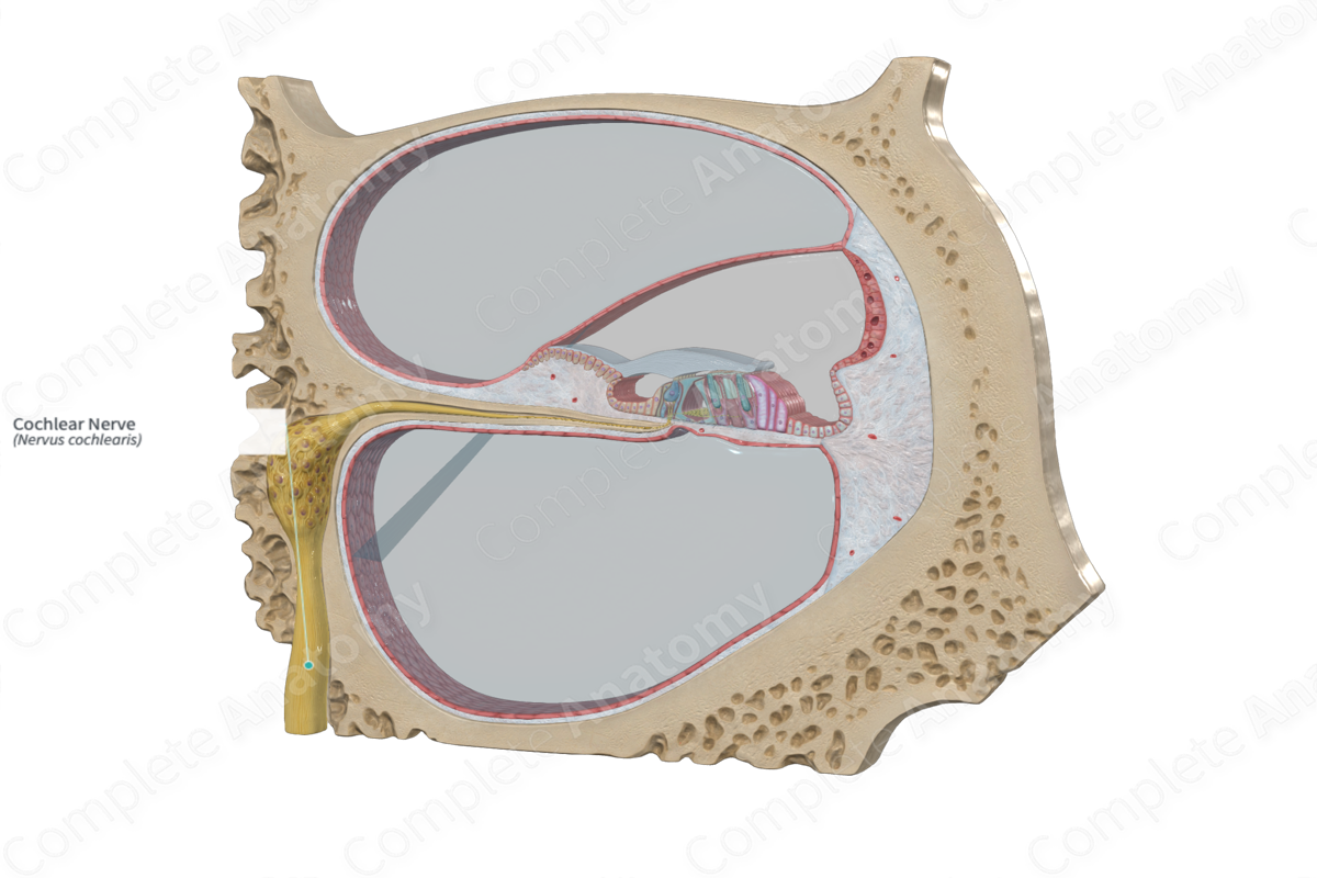
The cochlear nerve is the part of the vestibulocochlear nerve concerned with hearing, consisting of fibers that arise from the bipolar cells in the spiral ganglion and have their receptors in the spiral organ of the cochlea (Dorland, 2011).
Structure and/or Key Feature(s)
The cochlear nerve originates as one of two branches of the vestibulocochlear nerve, or the eighth cranial nerve (CNVIII), in the internal acoustic meatus. The cochlear nerve runs from the internal acoustic meatus laterally a short distance to the cochlea within the inner ear. The cochlear nerve is the sensory nerve responsible for hearing. It conveys special sense afferent fibers from the spiral organ of Corti to the cochlear nuclei within the pons and medulla of the brain.
The cochlear nerve is comprised of 30,000–40,000 afferent and efferent somatic nerve fibers. The diameter of these fibers varies, ranging from 1 to 11 μm. The afferent fibers possess myelinated axons, with bipolar cell bodies found in the spiral ganglion (in the modiolus). There are two distinguishable types of ganglion cells, type 1 (95%) and type 2 (5%). Each cell body emits a central process that protrudes toward the organ of Corti and a central process that projects back toward the cochlear nerve. The processes of type 1 cells are myelinated, while those of type 2 are not. From the modiolus, the peripheral branches of both cell types pass to the osseous spiral lamina and the type 1 branches are demyelinated here. From here, they pass via the habenula perforata of the spiral limbus to reach the organ of Corti and its mechanosensory hair cells (Moller, 2000).
At the base of each inner and outer hair cell, there is a cluster of nerve fibers forming a synaptic complex. This inner hair cell complex is comprised of the terminal afferent fibers of ten type 1 ganglion cells, with non-direct, lateral efferent contacts. The outer hair cell complex receives direct efferent contacts.
The dendrites of the afferent fibers do not run in parallel from the cell body to the hair cell; rather they are grouped into three different groups, depending on how they travel. These are the inner radial, basilar, and outer spiral fibers.
—The inner radial fibers make up the bulk of the afferent fibers and travel in a linear direction to reach the base of the inner hair cell.
—The basilar fibers run in a unique path, typically the length of five pillar cells, before deviating at an angle to form part of the afferent supply of the outer spiral bundle.
—The outer spiral bundle travels toward the base of the cochlea, with branches made of afferent and efferent fibers to supply the base of the outer hair cells.
The primary difference between the afferent nerve supply to the inner and outer hair cells is that a single inner hair cell is innervated by numerous afferent nerve fibers, whereas a single afferent nerve fiber innervates many outer hair cells.
Efferent cochlear fibers originate from the olivocochlear system located in the brainstem. They form the interganglionic spiral bundle in the modiolus. These efferent fibers are separated into the lateral and medial groups. The lateral efferent fibers are associated with the inner hair cells. They travel in the inner spiral bundle and synapse lateral to the afferents supplying the inner hair cell. They do not synapse directly with the base of the inner hair cell. In contrast, the medial efferent fibers are associated with the outer hair cells. These are myelinated and travel across the tunnel of Corti to synapse at the base of the outer hair cells. The overall efferent supply is not uniform throughout the cochlea. The inner row of the outer hair cells at the base of the cochlea has the richest supply. This is gradually reduced toward the apex of the cochlea and with the outer rows of the hair cells (Moller, 2000).
Function
The afferent fibers of the cochlear nerve relay signals generated from the hair cell back to the auditory processing centers in the brain. These synapses located at the inner hair cell provide information about the acoustic environment. In contrast, the lateral efferent fibers can alter the transmission of these afferent signals by their lateral input to the postsynaptic regions of these fibers.
Regarding the efferent cochlear fibers, the outer hair cells provide information about the acoustic environment. They possess a strong frequency discrimination, enabling them to alter the sensitivity of specific frequencies. This alteration of sensitivity and specificity is mediated via the medial efferent supply, which enables a change in the mechanical responses of the outer hair cells. These vestibular efferent fibers allow for higher centers to exert control of the activity of the cochlea, adapting it to the external auditory environment, in order to focus on particular sounds or to protect the auditory system.
List of Clinical Correlates
—Acoustic neuroma
References
Dorland, W. (2011) Dorland's Illustrated Medical Dictionary. 32nd edn. Philadelphia, USA: Elsevier Saunders.
Moller, A. R. (2000) Hearing: Its Physiology and Pathophysiology. Elsevier Science.
