Quick Facts
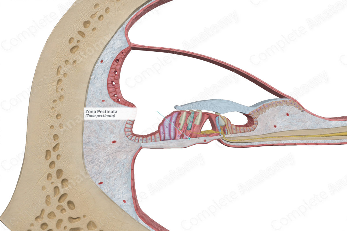
The zona pectinata is the outer two-thirds of the basilar membrane, running between the floor of the inner tunnel and the spiral ligament (Dorland, 2011).
Related parts of the anatomy
Structure and/or Key Feature(s)
The zona pectinata is one of the two regions of the basilar membrane, the other being the zona arcuata. The zona pectinata forms the outer part of the basilar membrane. It is trilaminar and thicker than the zona arcuata. The zona pectinata attaches from the base of the outer pillar cells and inserts into the basal (or spiral) crest, a triangular projection from the spiral ligament. On its insertion in the basal crest, the top and bottom layers of this trilaminar basilar membrane blend together.
Anatomical Relations
The zona pectinata forms the outermost part of the basilar membrane, which is interjacent between the scala tympani and the cochlear duct. The inferior surface of the zona pectinata is lined by periosteum, which, in turn, is lined with perilymphatic cells.
Function
The zona pectinata provides a sturdy foundation to the zona arcuata. Together, the two layers comprise a barrier which separates the cochlear duct and the scala tympani, as well as the endolymph and perilymph that occupy those canals. These two fluids have different ion concentrations involved in the transmission of vibrations. However, the zona arcuata is permeable to the perilymph of the scala tympani to an extent, thus allowing the spiral organ (of Corti) to bathe in perilymph (also referred to cortilymph in this region due to some minor alterations of its composition) (Standring, 2016).
References
Dorland, W. (2011) Dorland's Illustrated Medical Dictionary. 32nd edn. Philadelphia, USA: Elsevier Saunders.
Standring, S. (2016) Gray's Anatomy: The Anatomical Basis of Clinical Practice., 41st edition. Elsevier Limited.
