Quick Facts
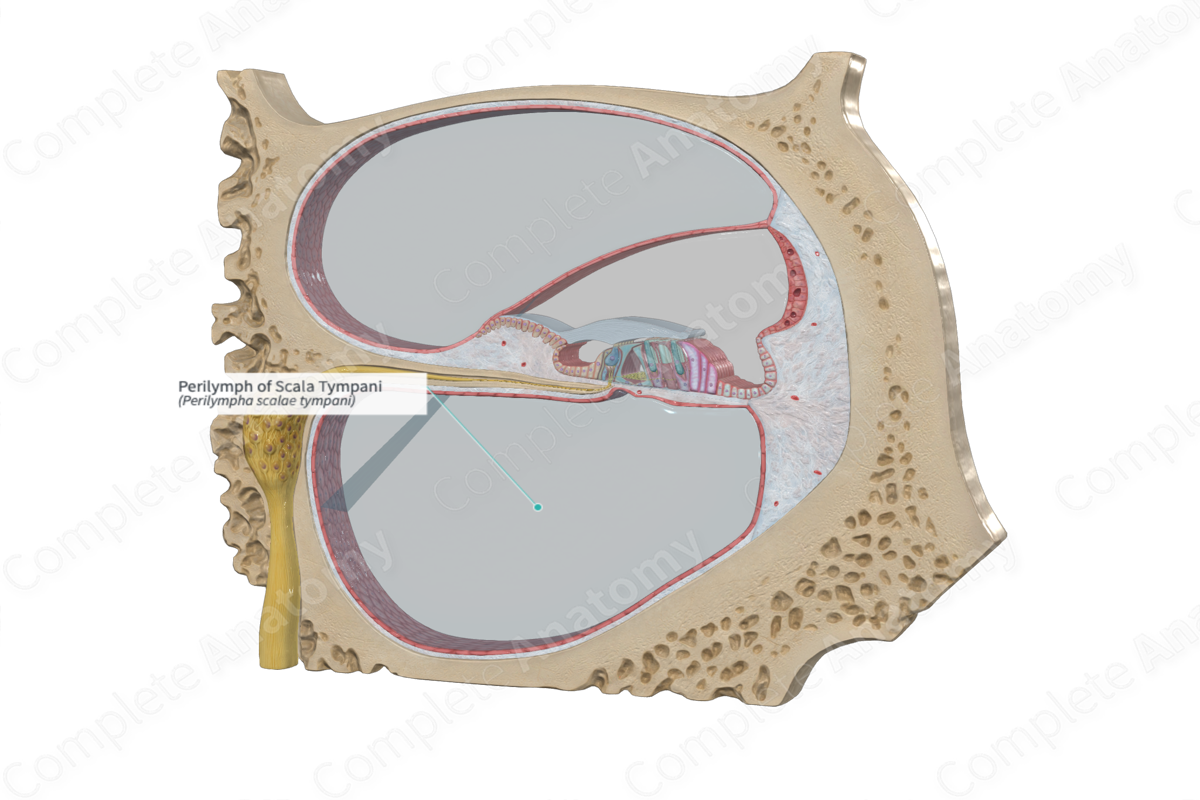
Perilymph is the fluid contained within the space separating the membranous labyrinth from the osseous labyrinth; it is entirely separate from the endolymph (Dorland, 2011).
Related parts of the anatomy
Structure and/or Key Feature(s)
The scala tympani is filled with perilymph, an extracellular fluid that is similar in consistency to that of cerebrospinal fluid, especially regarding the perilymph of the scala tympani. It receives some cerebrospinal through the cochlear canaliculus, which carries it from the subarachnoid spaces. Its dominant ion concentration is that of sodium.
Function
The perilymph is located between the bony labyrinth and the cochlear duct. It serves as a medium to transmit vibrations from the tympanic membrane (via the ossicles) to the endolymph in the cochlear duct, thus stimulating the spiral organ (of Corti). It also receives effluxing potassium released by the depolarization of the stereocilia.
The perilymph plays an important role in the sensory cell depolarization, as it receives the efflux of potassium from calcium-dependent channels in the stereocilia hair cells. It also contributes to the influx of calcium through specific channels located on the side and basal aspects of the hair cells.
References
Dorland, W. (2011) Dorland's Illustrated Medical Dictionary. 32nd edn. Philadelphia, USA: Elsevier Saunders.
Standring, S. (2016) Gray's Anatomy: The Anatomical Basis of Clinical Practice., 41st edition. Elsevier Limited.
