Quick Facts
The perilymphatic cells maintain the balance of ionic contents in the perilymph and endolymph.
Related parts of the anatomy
Structure and/or Key Feature(s)
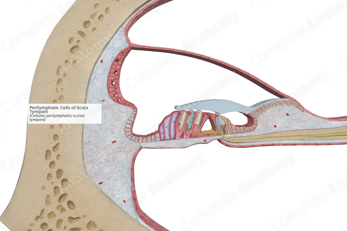
The inner aspect of the bony labyrinth is lined by perilymphatic cells with interspersed extracellular fibers. These perilymphatic cells have varying cell morphology based on their location in the bony labyrinth.
The perilymphatic cells lining the periosteum are generally very flat, resembling a squamous epithelium. They contain tight junctions, which creates a diffusion barrier between it and the endolymph in the cochlear duct. Perilymphatic cells on the basilar membrane facing into the scala tympani are cuboidal in shape.
In the narrower areas of the cochlea, such as where the scala vestibuli approaching the helicotrema, the perilymphatic cells have a different form. They have a stellate or reticular (cross-linked in a network fashion) in appearance and may have thin cytoplasmic projections extending across the space (Standring, 2016).
Function
The perilymphatic cells maintain the balance of ionic contents in the perilymph and endolymph.
List of Clinical Correlates
—Meniere's disease
References
Standring, S. (2016) Gray's Anatomy: The Anatomical Basis of Clinical Practice., 41st edition. Elsevier Limited.
