Quick Facts
Origin: Proper hepatic artery.
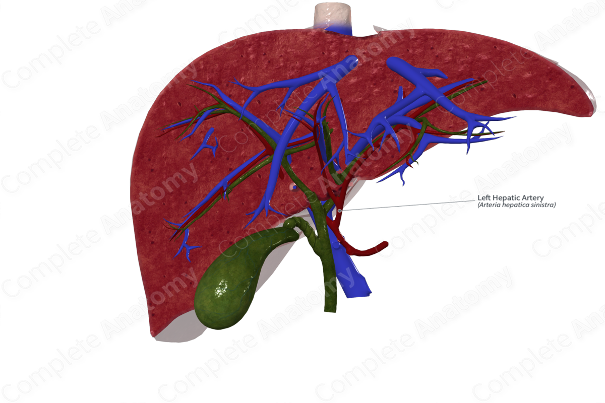
Course: Enters the porta hepatis of the liver.
Branches: Left artery of caudate lobe and medial and lateral segmental arteries.
Supplied Structures: Left “functional” lobe of the liver that is composed of segments I (caudate lobe), II, III, and IV.

Origin
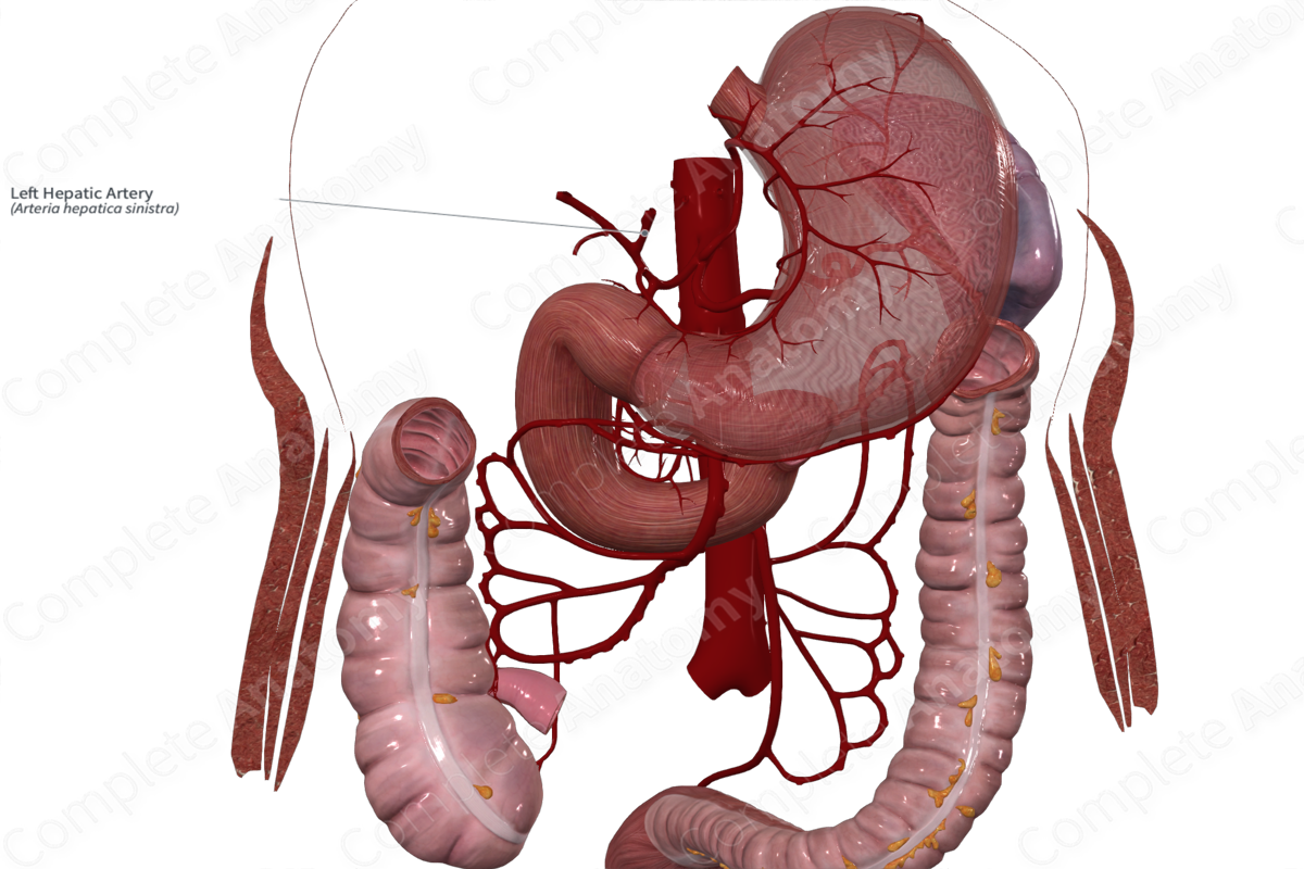
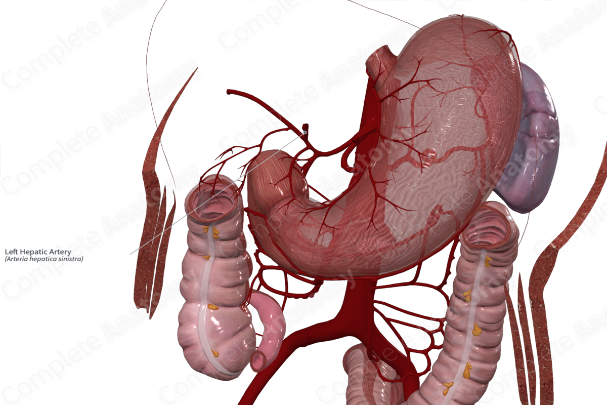
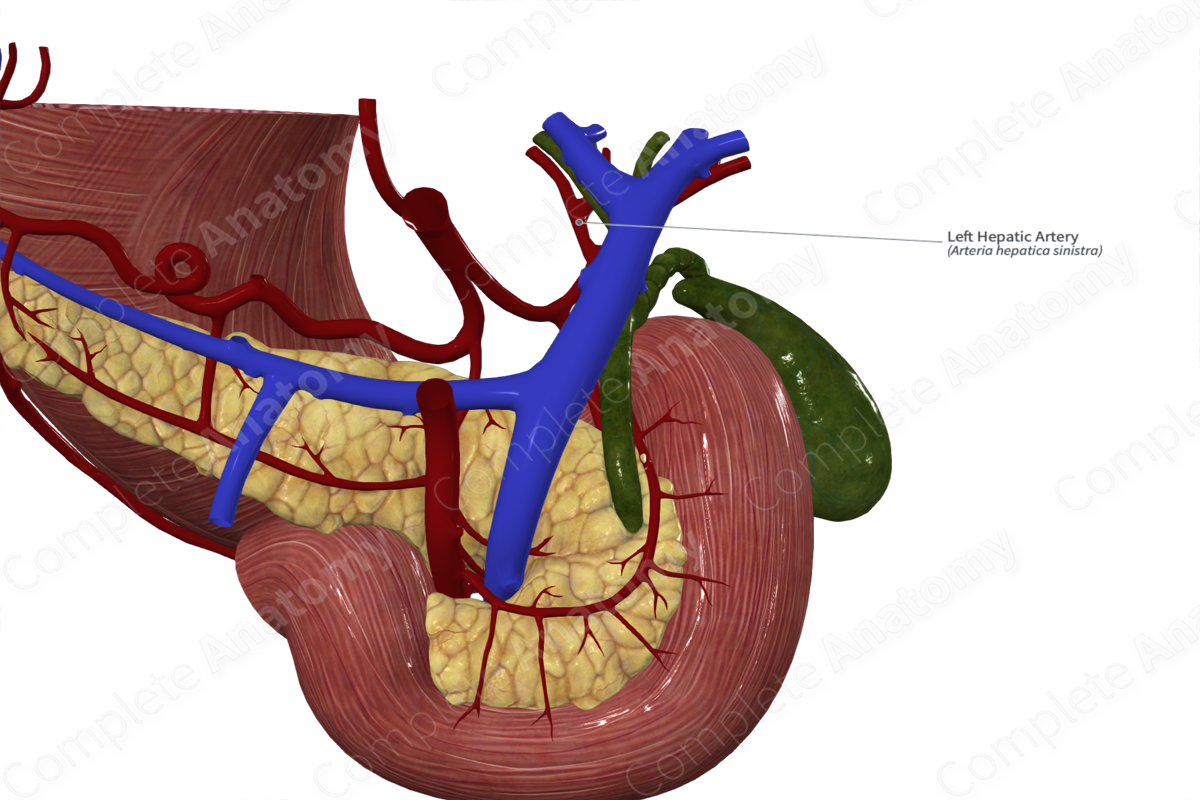
The left hepatic artery arises from the proper hepatic artery.
Course
The proper hepatic artery ascends nearly vertically within the hepatoduodenal ligament. It travels as part of the portal triad, to the left of the common bile duct and anterior to the portal vein. At a variable position along its course, it branches into right and left hepatic branches. Most often this bifurcation occurs just inferior to the porta hepatis. After branching the left hepatic artery angles to the left and enters the liver parenchyma. Usually within the liver parenchyma the left hepatic artery arches over the portal vein (arch of the left hepatic artery), a defining feature that helps distinguish it on contrast radiographs.
Anatomical variation in the disposition of the arteries to the liver occurs in about one third of individuals (Covey et al., 2002; Saba and Mallarini, 2011).
Branches
The left hepatic artery usually gives off a left artery of the caudate lobe, thus, forming an anastomosis between the left and right hepatic arteries via the right artery of the caudate lobe. The left hepatic artery also gives rise to medial and lateral segmental arteries.
Supplied Structures
The left hepatic artery supplies the left “physiological” lobe of the liver composed of segments I (caudate lobe), II, III, and IV. Segment I usually exhibits shared vascularization from the left and right hepatic arteries.
References
Covey, A. M., Brody, L. A., Maluccio, M. A., Getrajdman, G. I. and Brown, K. T. (2002) 'Variant hepatic arterial anatomy revisited: digital subtraction angiography performed in 600 patients', Radiology, 224(2), pp. 542-7.
Saba, L. and Mallarini, G. (2011) 'Anatomic variations of arterial liver vascularization: an analysis by using MDCTA', Surg Radiol Anat, 33(7), pp. 559-68.
Learn more about this topic from other Elsevier products

