Quick Facts
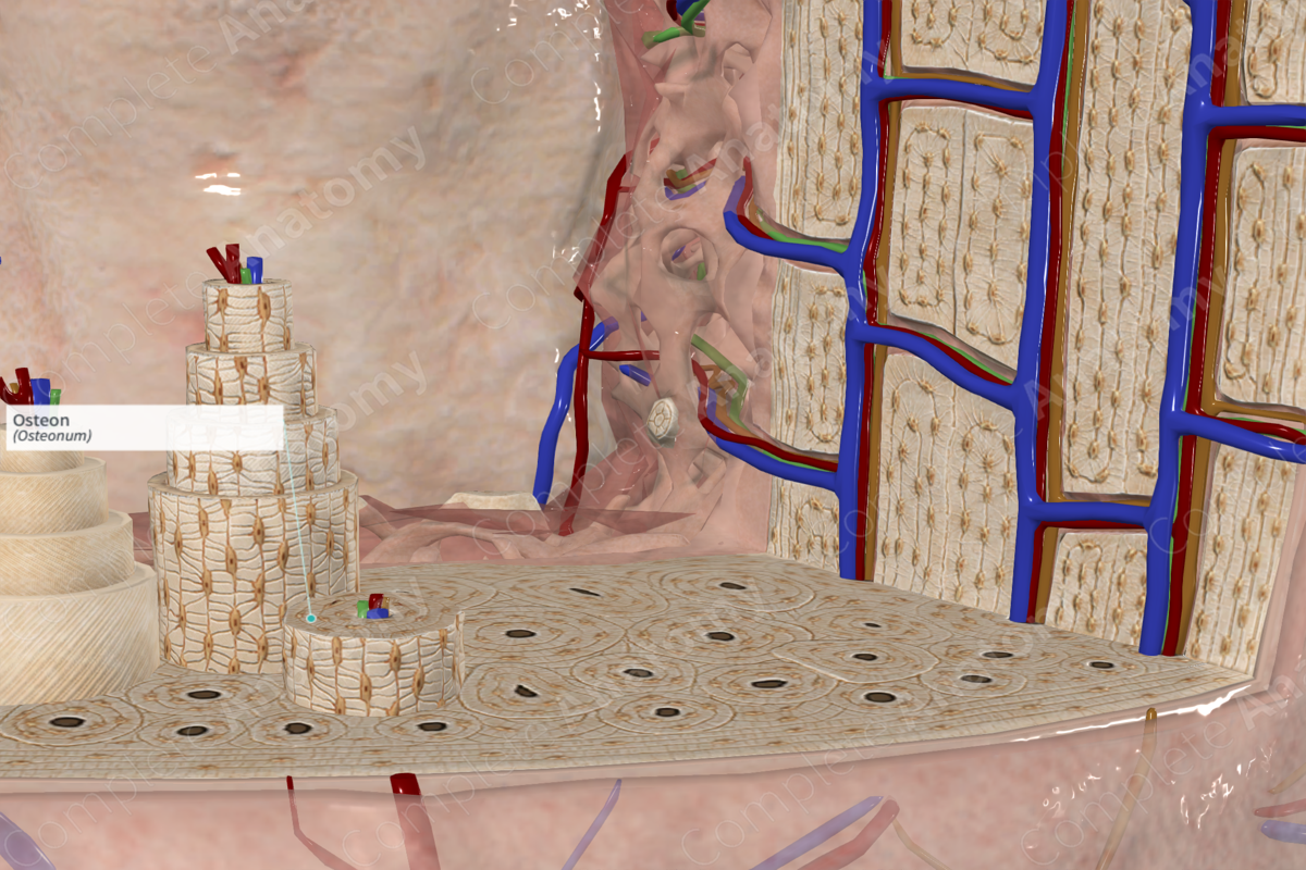
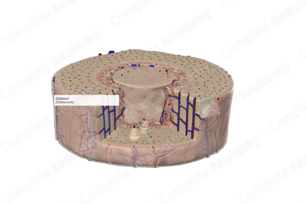
The osteon is the basic unit of structure of compact bone, comprising a central (haversian) canal and its concentrically arranged lamellae, of which there may be 4 to 20, each 3 to 7 μm thick, in a single (haversian) system; such units are directed mainly in the long axis of the bone (Dorland, 2011).
Structure/Morphology
Osteons are the cylindrical, functional units of bone. In adults, osteons mainly form in existing compact bone through the continuous process of bone remodeling. In long bones, osteons form parallel to each other and parallel to the long axis of the bone and, thus, appear round in cross-sectional anatomy. Osteons can be identified by their central nutrient (or Haversian) canal, which provides a channel for vasculature, nerves, and lymphatics to travel. A population of cells, the osteoblasts, produce 5–20 rings of lamellar bone that radiate outward from nutrient canal (Standring, 2016). Haversian canals can communicate with each other via Volkmann’s canals, which run obliquely to the axes of the osteons.
Osteocytes are formed when actively secreting osteoblasts become surrounded by osteoid and become trapped in spaces called lacunae. The osteocytes, in their lacunae, are parallel to the lamellar rings. These lacunae are connected via small channels called canaliculi. The canaliculi radiate from lacunae in a perpendicular orientation. Osteocytes extend their slender cellular process through these spaces to communicate with each other. This facilitates cell-cell communication via gap junctions (Ross and Pawlina, 2006).
Anatomical Relations
In cortical bone interstitial lamellae are found in irregular orientations between osteons. They are separated from osteons by a thin layer called the cement line. The internal and external circumferential lamellae are the inner and external limits for osteons in cortical bone.
Function
Osteons serve as the functional unit of bone. Their thick lamellar rings provide mechanical support and strength to bone. They protect the vascular supply via the nutrient canal. Osteons are constantly remodeling and respond to the biomechanical stresses that they undergo (Standring, 2016).
References
Dorland, W. (2011) Dorland's Illustrated Medical Dictionary. 32nd edn. Philadelphia, USA: Elsevier Saunders.
Ross, M. H. and Pawlina, W. (2006) Histology: A text and atlas. Lippincott Williams & Wilkins.
Standring, S. (2016) Gray's Anatomy: The Anatomical Basis of Clinical Practice. Gray's Anatomy Series: Elsevier Limited.
