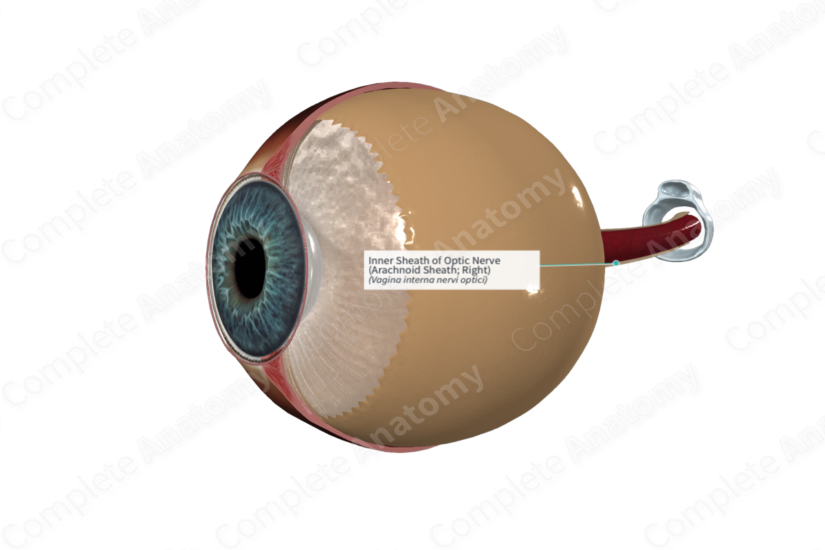
Inner Sheath of Optic Nerve (Arachnoid Sheath; Right)
Vagina interna nervi optici
Read moreQuick Facts
The inner sheath of optic nerve is the internal sheath of the optic nerve, continuous with the pia mater and arachnoid mater (Dorland, 2011).


Structure and/or Key Feature(s)
The eye and optic nerve are outgrowths of the brain and as such carry the three layers of meningeal coverings with them. They are called collectively, the optic sheath. Of these, the inner two layers, arachnoid and pia, are referred to collectively as the inner or internal sheath of the optic nerve.
The outermost layer of the inner sheath of the optic nerve is continuous with the arachnoid mater of the brain. It is referred to as the arachnoid sheath of the inner sheath of the optic nerve. As the optic nerve enters the eyeball, the arachnoid sheath becomes continuous with the choroid of the eyeball (Mukherjee, El-Dairi and Bhatti, 2013).
Anatomical Relations
The arachnoid sheath is sandwiched between the external dural sheath, separated from it by a subdural space, and the internal pial sheath, separated from it by a subarachnoid space. The subarachnoid space is filled with cerebrospinal fluid continuous with that of the brain. The sheaths end as the optic nerve enters the eyeball to form the optic nerve head.
Function
Contrary to popular notion, the optic nerve is not a peripheral nerve but a white matter tract of the central nervous system. As such it is covered, and partially supported by the same three meningeal sheaths and their fluid filled separating spaces as the brain. These perform the same support functions for the optic nerve (Mukherjee, El-Dairi and Bhatti, 2013).
References
Dorland, W. (2011) Dorland's Illustrated Medical Dictionary. 32nd edn. Philadelphia, USA: Elsevier Saunders.
Mukherjee, N., El-Dairi, M. A. and Bhatti, M. T. (2013) 'Optic Nerve Sheath Fenestration—Indications and Techniques', US Ophthal. Rev., 6(2), pp. 125-131.
