Quick Facts
Origin: From the venules inside the retina.
Course: Accompanies central retinal artery in the optic nerve.
Tributaries: Superior and inferior nasal and temporal branches.
Drainage: Retina.
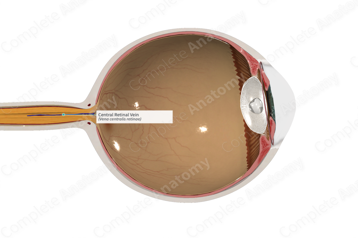
Origin
The central retinal vein arises from the union of the superior and inferior nasal and temporal venules in the retina.
Course
Within the eyeball, the intraocular part of the central retina vein does not share a similar distribution with the arteries. Arteries, which lie more superficially, can often cross over the veins. In individuals with severe hypertension, the arteries press on the veins causing dilations of the veins.
The extraocular part of the central retinal vein accompanies the central retinal artery within the optic nerve. It is short and drains either directly into the cavernous sinus, or into the superior ophthalmic vein.
Tributaries
The central retinal vein receives the superior and inferior nasal and temporal venules, which each drain a quadrant of the retina.
Structures drained
The central retinal vein is responsible for draining the inner layers of the retina of the eyeball including the outer plexiform layer, the inner nuclear layer, the inner plexiform layer, and the ganglion cell layer.
List of Clinical Correlates
—“Nicking” of central retinal veins as an indication of hypertension observable upon ophthalmoscopic examination
