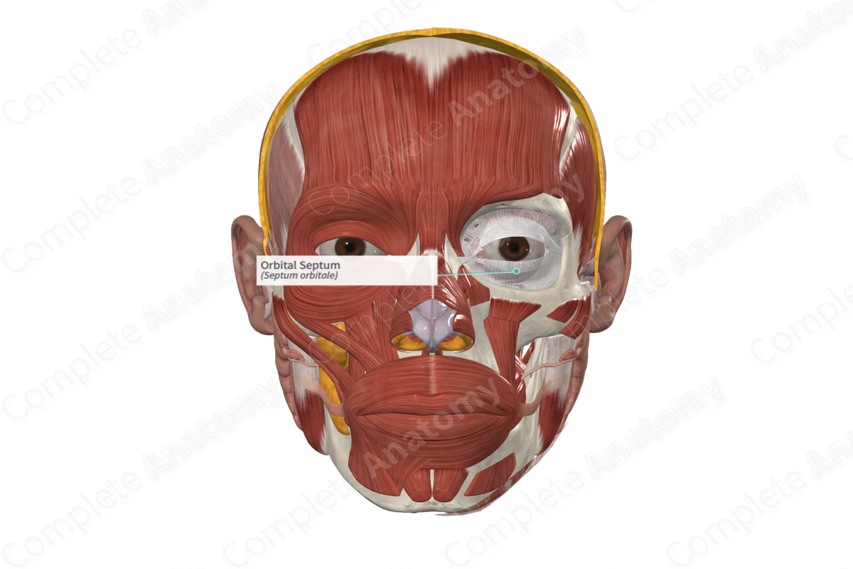
Structure
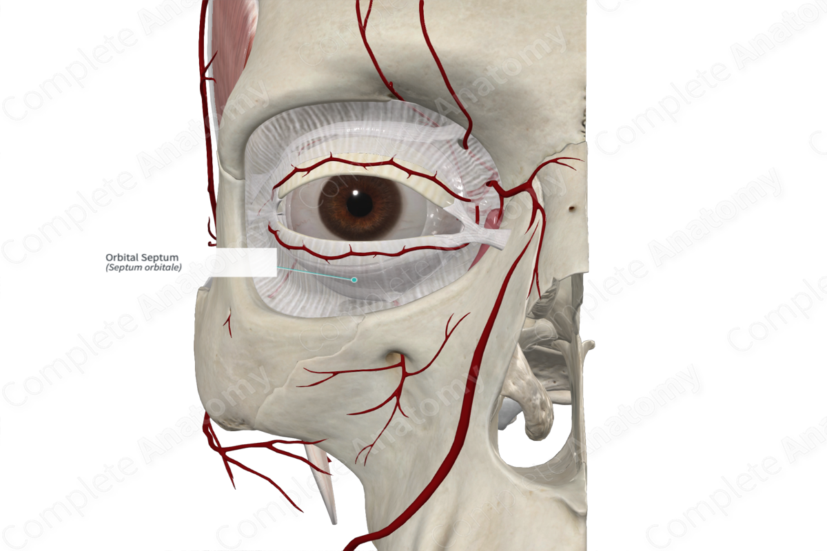
The orbital septum is a weak membranous sheet that attaches to the rim of the orbit. It is continuous with the periosteum and is thickest laterally, where it lies anterior to the lateral palpebral ligament.
Related parts of the anatomy
Anatomical Relations
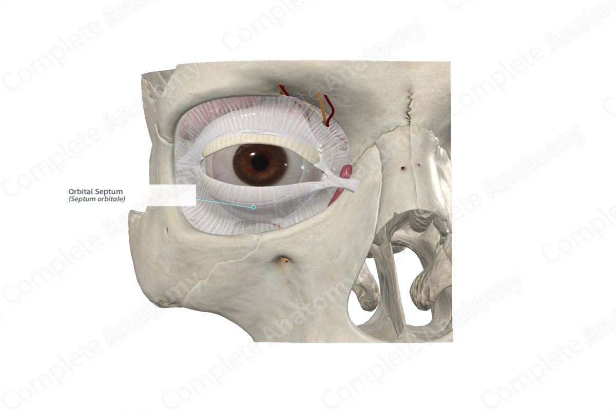
The orbital septum extends into the eyelid where it blends with the tarsal plates and the superficial lamella of the levator palpebrae superioris muscle.
Laterally, the orbital septum lies anterior to the lateral palpebral ligament, whereas medially, the orbital septum passes posteriorly to the medial palpebral ligament and the nasolacrimal sac.
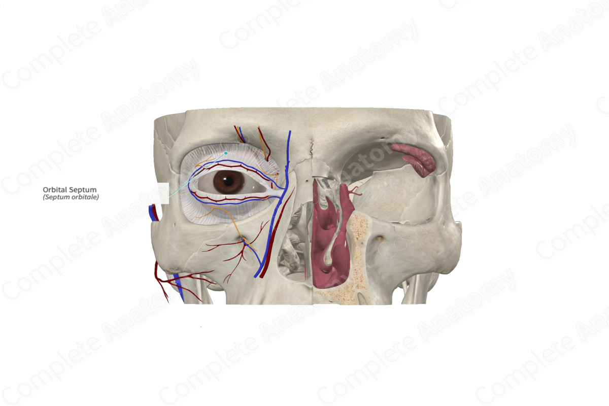
The septum is pierced by the extraocular muscle, levator palpebrae superioris, and fibrous extensions of the sheaths of inferior oblique and inferior rectus muscles. It is also pierced by nerves and vessels that pass from the orbit to the face and scalp.
Function
When the eyes are closed, the orbital septum and tarsal plates provide complete coverage of the eyeball. The septum also acts to keep the eyelids stable as the eye moves.
List of Clinical Correlates
—Orbital cellulitis
—Periorbital cellulitis
—Age-related eyelid defects
Learn more about this topic from other Elsevier products

