Structure
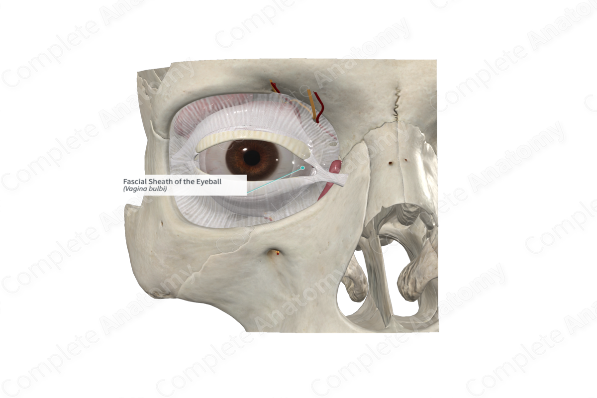
The fascial sheath of the eyeball surrounds the eye, extending from the optic nerve to the corneoscleral junction. The fascial sheath is thickened anteriorly at the attachment of the recti muscles, thus, forming a fascial ring.


Related parts of the anatomy
Anatomical Relations
At the posterior aspect, the fascial sheath is traversed by ciliary vessels and nerves. It is also perforated by the tendons of the extraocular muscles, where it is reflected on to each muscle as a sheath. The reflection onto the superior oblique muscle extends to the trochlea, whereas the sheaths of the four rectus muscles are thick anteriorly but posteriorly form a thin perimysium.
These fascial sheaths around the extraocular muscles give off important expansions to the walls of the orbit, that limits the action of the medial and lateral rectus muscles. These expansions are named the medial and lateral check ligaments.
The sheath around the inferior rectus muscle is also thickened. It acts as a suspensory ligament of the eye due to it being continuous with the sheaths of the medial and lateral rectus muscles which are attached to the medial and lateral check ligaments.
Function
The fascial sheath of the eyeball separates the eyeball from the surrounding orbital fat.
List of Clinical Correlates
—Sub-Tenon’s anesthesia
Learn more about this topic from other Elsevier products

