Structure
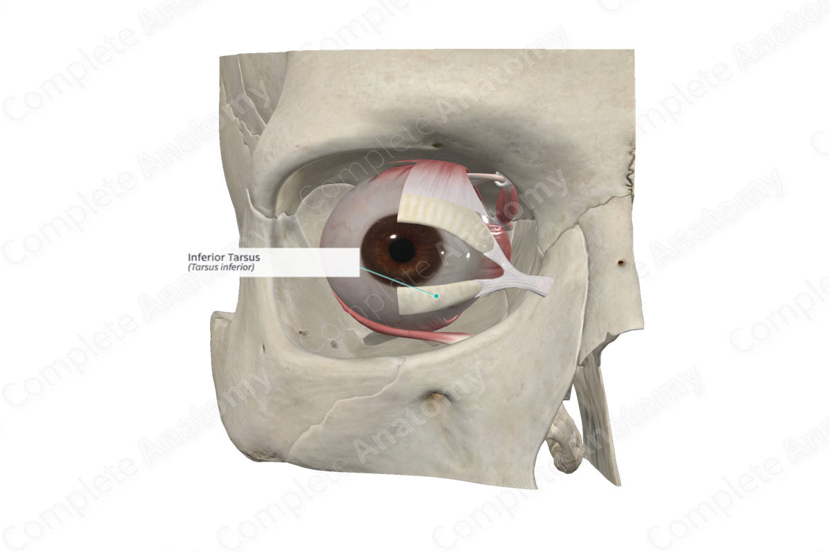
The superior and inferior eyelids contain a thin, elongated plate composed of tough collagenous tissue called the tarsal plate. Each plate is convex, conforming to the shape of the anterior surface of the eyeball. It has a free straight ciliary border adjacent to the hair follicles of the eyelashes, and a convex orbital border that is attached to the orbital septum (palpebral fascia). The inferior tarsus is the smallest of the two tarsal plates. It is approximately 2.5 cm long and 4 mm high (Standring, 2016).
Related parts of the anatomy
Anatomical Relations
The free ciliary margins of the tarsal plates end approximately 2 mm from the margin of the eyelid and runs parallel to it.
The orbital margins of the tarsal plates are bound to margins of the orbital septum and to the lateral and medial palpebral ligaments on each side. The medial palpebral (canthic) ligament passes from the medial ends of both tarsal plates and insets into the anterior lacrimal crest and the frontal process of the maxilla (bone forming part of the orbital cavity). As it inserts into the tarsal plates, the ligament splits to surround the lacrimal canaliculi. The lateral palpebral ligament, on the other hand, is poorly developed. It passes from the lateral edges of the tarsal plates to the zygomatic bone that forms part of the orbital margin.
Each tarsal plate has associated smooth muscle forming the superior and inferior tarsal muscles, which aids in elevating and depressing the upper and lower eyelids, respectively.
Function
The superior and inferior tarsal plates provide support to the eyelids and determine the shape of each lid.
References
Standring, S. (2016) Gray's Anatomy: The Anatomical Basis of Clinical Practice, 41st ed. Elsevier Limited.
