Morphology/Structure
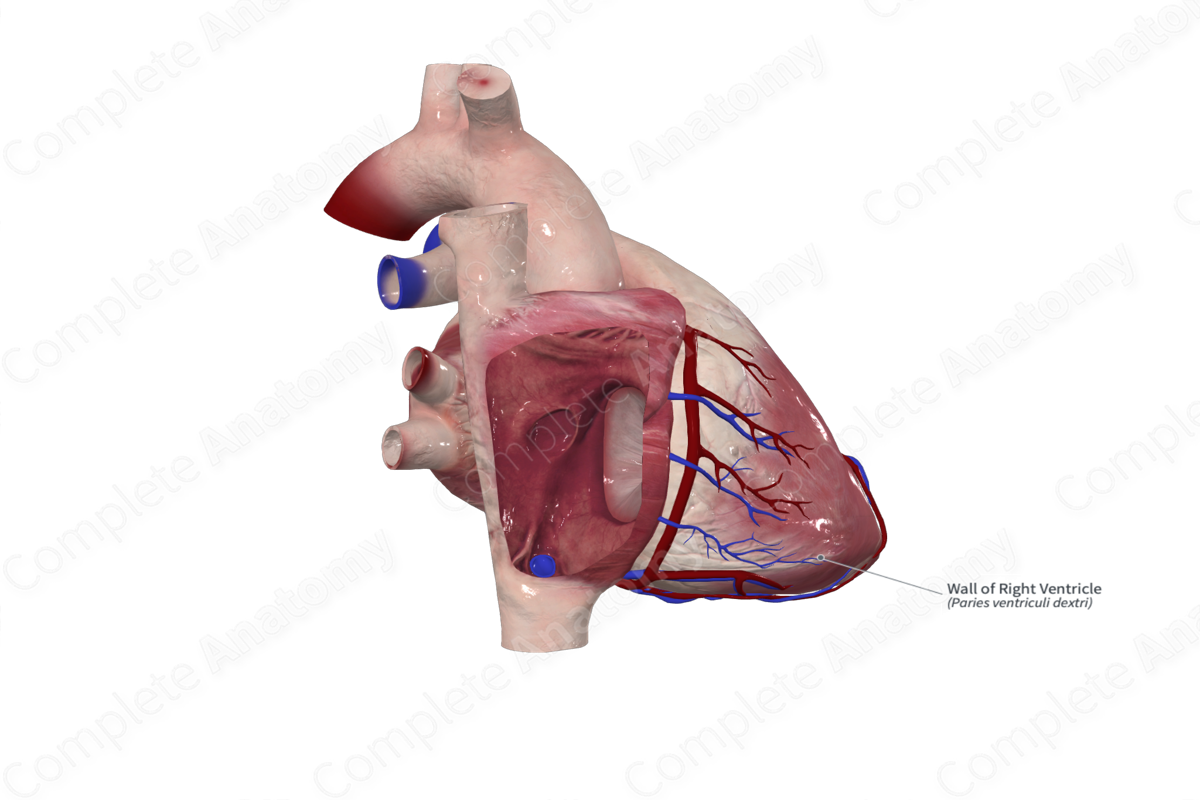
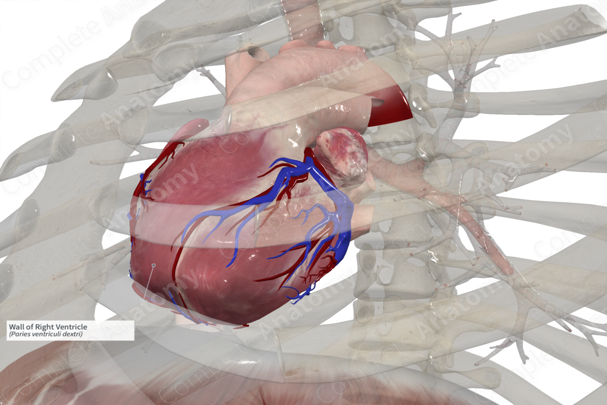
The right ventricle forms the majority of the anterior (sternocostal) surface of the heart and extends from the right atrioventricular valve almost to the apex of the heart.
The thickness of the right ventricular wall is only one third that of the left ventricle and is approximately 3–5 mm thick. Due to the dominance of the left ventricle, which forms the majority of the apex of the heart, the interventricular septum bulges into the right ventricle, giving it a crescent shape when viewed on a transverse cross section.
It has a smooth-walled outflow tract called the infundibulum (conus arteriosus) which extends superiorly and to the left as far as the pulmonary orifice. The majority of the ventricular walls contain ridges called the trabeculae carneae, which are highly varied in appearance. Additionally, the papillary muscles are muscular bundles that extend from the ventricular walls and end in thin collagenous cords, the chordae tendineae, which in turn insert into the leaflets of the right atrioventricular valve (or tricuspid valve).
One particular trabecula, the septomarginal trabecula, connects to the anterior papillary muscle and contains the right bundle of the atrioventricular bundle. It's an important component in the conducting system of the heart, where it aids in coordinated ventricular contraction.
Key Features/Anatomical Relations
The right ventricle is located inferior to the right atrium and is separated from the left ventricle by the interventricular septum.
Externally, the anterior surface of the left and right ventricles is divided by the anterior interventricular sulcus containing the anterior interventricular artery and the great cardiac vein.
On its inferior (diaphragmatic) surface, the left and right ventricles are divided by the posterior interventricular sulcus which contains the inferior interventricular branches (or posterior descending artery) and middle cardiac vein.
The coronary sulcus (atrioventricular sulcus) divides the atria above from the ventricles below and contains the main limbs of the coronary vessels. However, the sulci and vessels are often obscured with adipose tissue.
Function
During right atrial contraction (atrial systole), the right ventricle is relaxed (ventricular diastole), allowing the deoxygenated blood to enter the ventricle without resistance. As the right atrium relaxes and refills (atrial diastole), the right ventricle contracts (ventricular systole), forcing the deoxygenated blood through the pulmonary valve and into the pulmonary trunk for transport to the lungs.
Learn more about this topic from other Elsevier products

