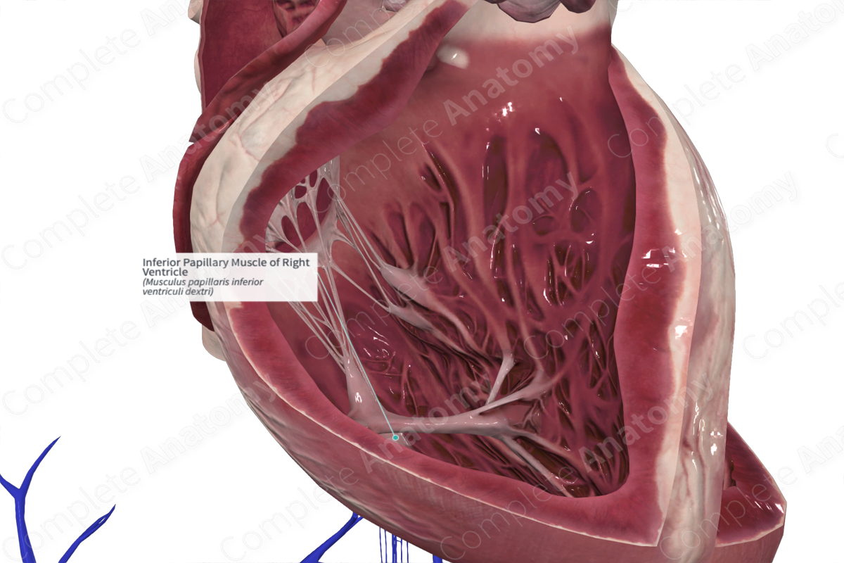
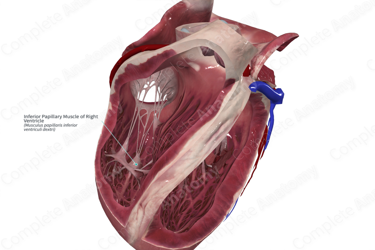
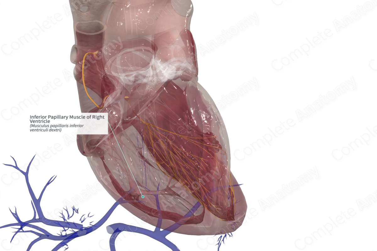
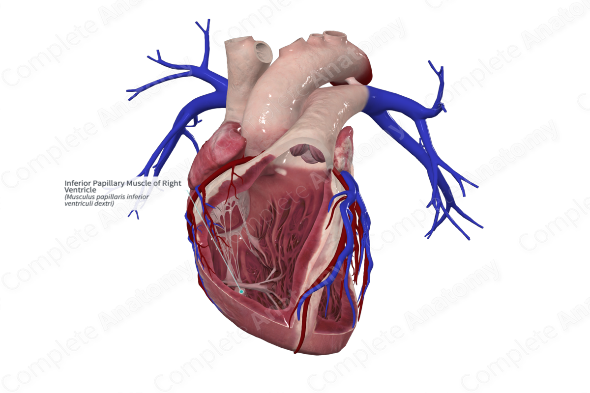
Inferior Papillary Muscle of Right Ventricle
Musculus papillaris inferior ventriculi dextri
Read moreMorphology/Structure
The inferior papillary muscle is one of the three papillary muscles found in the right ventricle. The other two papillary muscles are the larger anterior and the smaller septal papillary muscles. Each papillary muscle is a conical muscular bundle that extends from the ventricular wall and to the chordae tendineae. The chordae tendineae in turn insert into the leaflets of the right atrioventricular valve (or tricuspid valve). The chordae tendineae typically attach to the apical one third of the papillary muscles, but occasionally, attach to to their base.
Key Features/Anatomical Relations
The inferior papillary muscle is attached to the inferior and septal leaflets inferior to the inferoseptal commissure. It may be bifid or even trifid.
Function
During atrial contraction, the papillary muscles are relaxed, and the valve is open, avoiding any resistance to the movement of blood from the atrium to the ventricle. The papillary muscles contract just before ventricular systole. During ventricular contraction the valve leaflets close. This is achieved by papillary muscle contraction, thus pulling on the chordae tendineae and valve leaflets. This tension prevents the leaflets swinging backwards into the atria. The tension in the papillary muscles remains until the next atrial contraction.
The pulmonary valve does not require papillary muscles, as their inherent semilunar shape lends them the stability required to prevent retrograde blood flow and the extension of the pulmonary valve leaflets into the pulmonary trunk.
Learn more about this topic from other Elsevier products
Right Ventricular Papillary Muscle

