Quick Facts
Location: Vertebral column.
Bone Type: Irregular bone.
Key Features: Median and lateral parts, apex, base, sacral cornu, and sacral tuberosity.
Articulates With: Fifth lumbar vertebra and coccyx.
Arterial Supply: Iliolumbar, lateral sacral, and median sacral arteries.
Key Features & Anatomical Relations
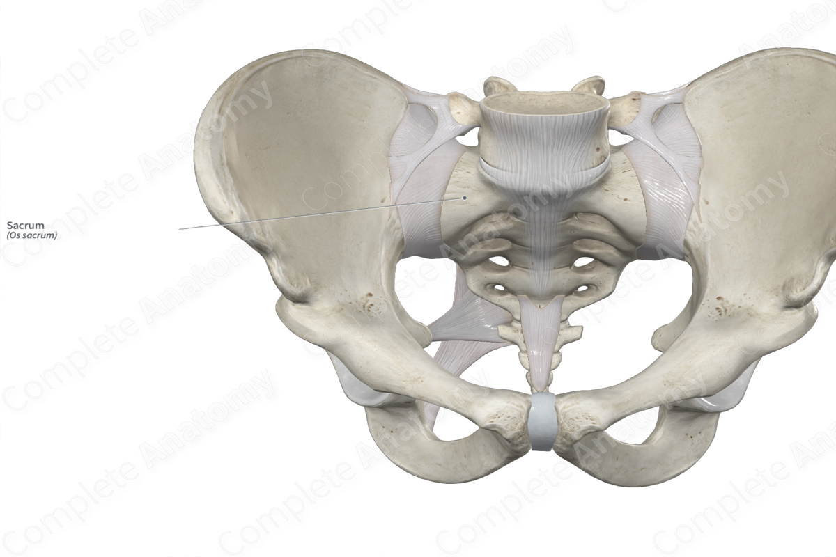
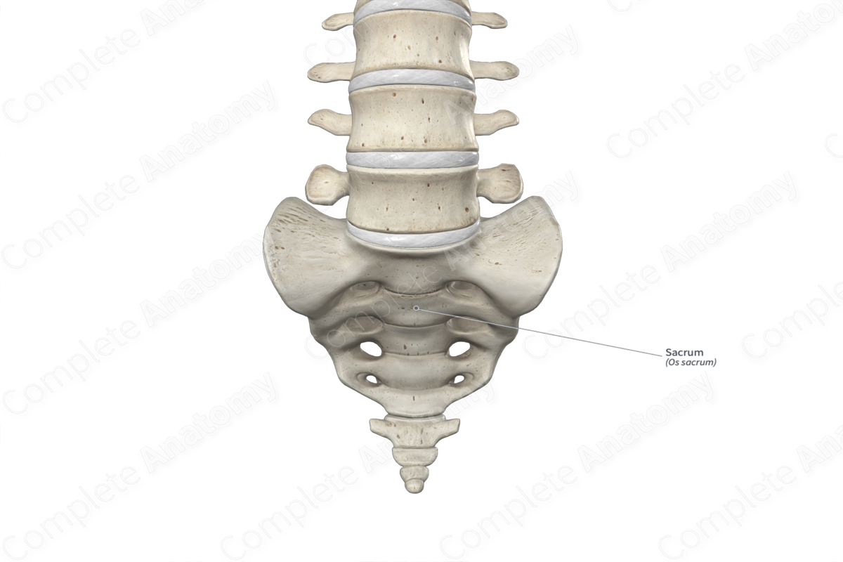
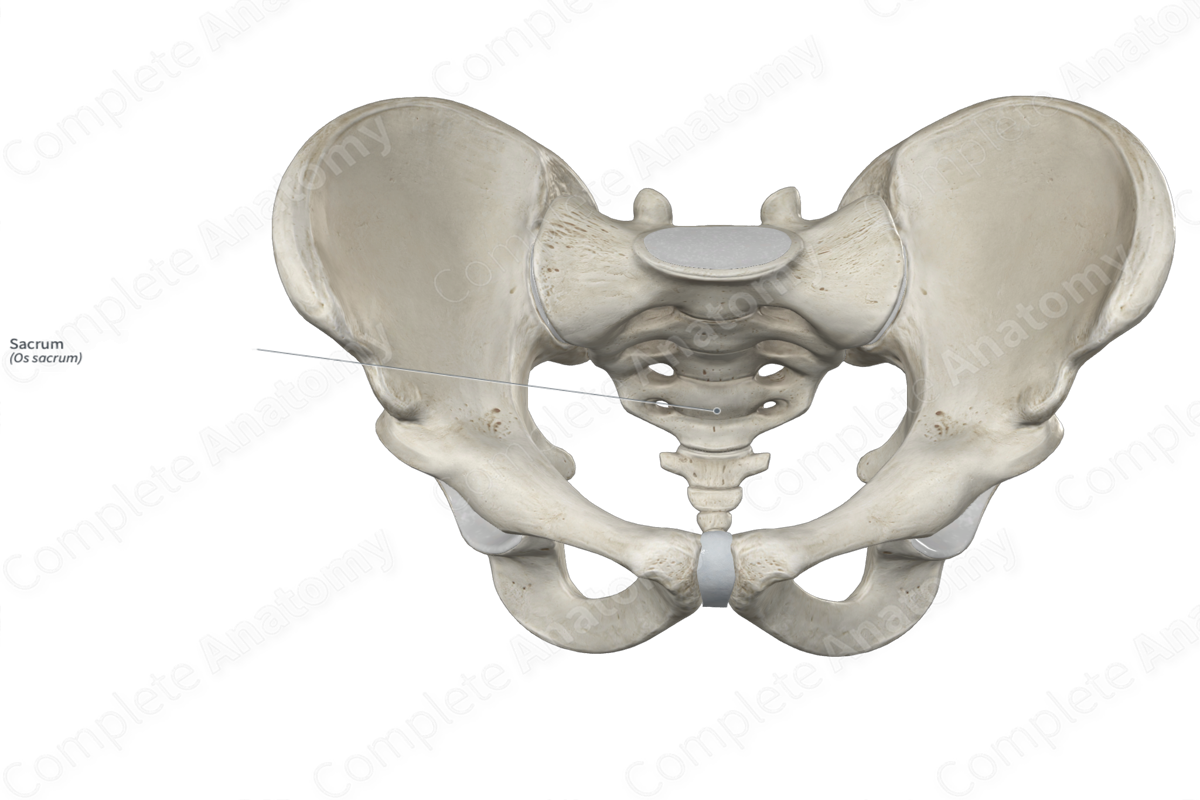
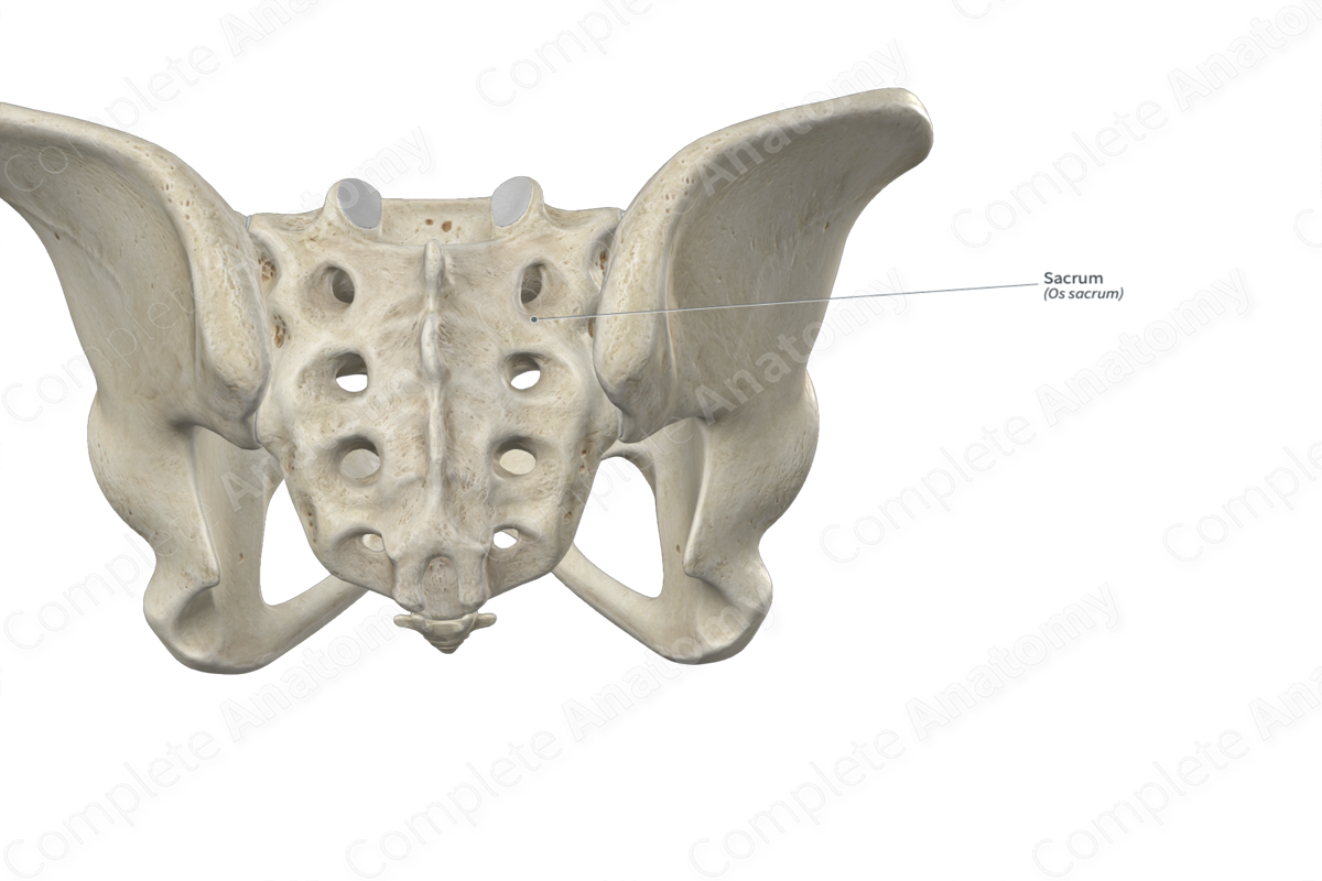
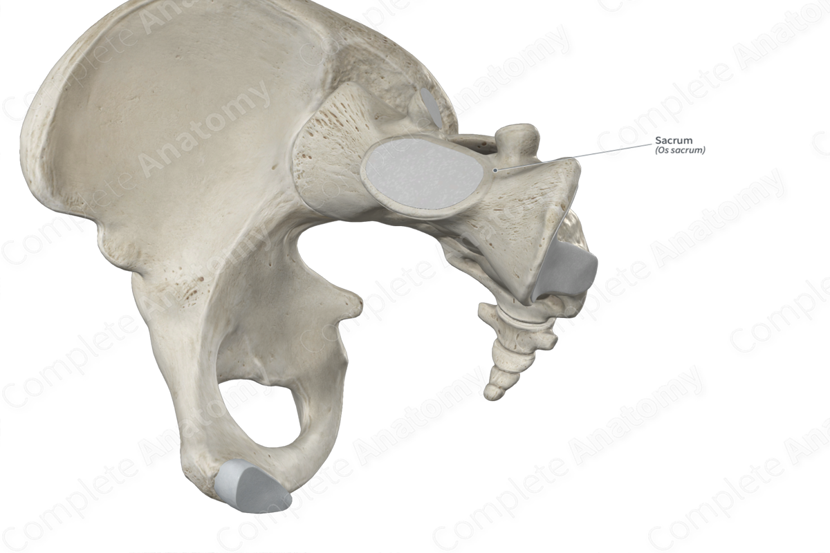
The sacrum is one of the five regional groups of the vertebral column. It is triangular, classified as an irregular bone, and includes the following bony features:
- parts: median and lateral parts;
- surfaces: apex, base, and pelvic, dorsal, and lateral surfaces;
- landmarks: ala, sacral cornu, sacral tuberosity, and median, intermediate, and lateral sacral crests.
More information regarding these and other bony features can be found in the Parts, Surfaces, and Landmarks tabs for this bone.
The sacrum is located:
- posterior to the pelvic cavity;
- medial to the iliac bones;
- superior to the coccyx;
- inferior to the fifth lumbar vertebra.
It contributes to the formation of the pelvic girdle, and it articulates with:
- iliac bones at the sacroiliac joints;
- coccyx at the sacrococcygeal joint;
- fifth lumbar vertebra at the lumbosacral joint.
Ossification
Ossification of the sacrum occurs at each of the five sacral vertebrae that unite to form the sacrum. Ossification of each of these vertebrae occurs at five ossification centers, these are found in the:
- body, which appears in utero during the third to fifth months;
- right half of the vertebral arch, which appears in utero during the third to fifth months;
- left half of the vertebral arch, which appears in utero during the third to fifth months;
- right transverse tubercle of each sacral vertebra, which collectively form the right lateral sacral crest, and appears in utero during the sixth to eighth months;
- left transverse tubercle of each sacral vertebra, which collectively form the left lateral sacral crest, and appears in utero during the sixth to eighth months.
In each sacral vertebra, their ossification centers fuse with each other during the second to eighth years. The five sacral vertebrae begin to fuse with each other during early adolescence and this continues all the way up to early adulthood (Standring, 2016).
Variations
In some individuals:
- the fifth lumbar vertebra may be fused with the sacrum, i.e., sacralization of the fifth lumbar vertebra;
- the first coccygeal vertebra may be fused with the sacrum, i.e., sacralization of the first coccygeal vertebra;
- the first sacral vertebra may be unfused with the rest of the sacrum and have similar characteristic features to those of the lumbar vertebrae, i.e., lumbarization of the first sacral vertebra (Tubbs, Shoja and Loukas, 2016).
The sacrum also displays sexual dimorphism, where the sacrum tends to be less curved, shorter, and wider in females.
Surface Anatomy
- The dorsal surface of the sacrum can be palpated between the right and left iliac bones.
- The median sacral crest can be palpated along the midline of the dorsal surface, in line with the spinous processes of the lumbar vertebrae.
- The sacral hiatus can be palpated as the flat or hollow area that is found at the inferior end of the median sacral crest.
List of Clinical Correlates
- Fracture of sacrum
- Sacrococcygeal teratoma
- Sacroiliac (SI) joint dysfunction
References
Standring, S. (2016) Gray's Anatomy: The Anatomical Basis of Clinical Practice. Gray's Anatomy Series 41st edn.: Elsevier Limited.
Tubbs, R. S., Shoja, M. M. and Loukas, M. (2016) Bergman's Comprehensive Encyclopedia of Human Anatomic Variation. Wiley.
Learn more about this topic from other Elsevier products

Sacrum muscle energy treatment: Video and Anatomy

Master Sacrum muscle energy treatment: anatomy, function, and structure. High-yield review with labeled diagrams, videos, and practice quizzes.
