Quick Facts
Location: Viscerocranium.
Bone Type: Irregular bone.
Key Features: Zygomatic, frontal, alveolar, and palatine processes, body, canine fossa, maxillary tuberosity, and anterior lacrimal crest.
Articulates With: Opposite maxilla, ethmoid, frontal, lacrimal, palatine, nasal, and zygomatic bones, vomer, and inferior nasal concha.
Arterial Supply: Facial and maxillary arteries.
Related parts of the anatomy
Key Features & Anatomical Relations
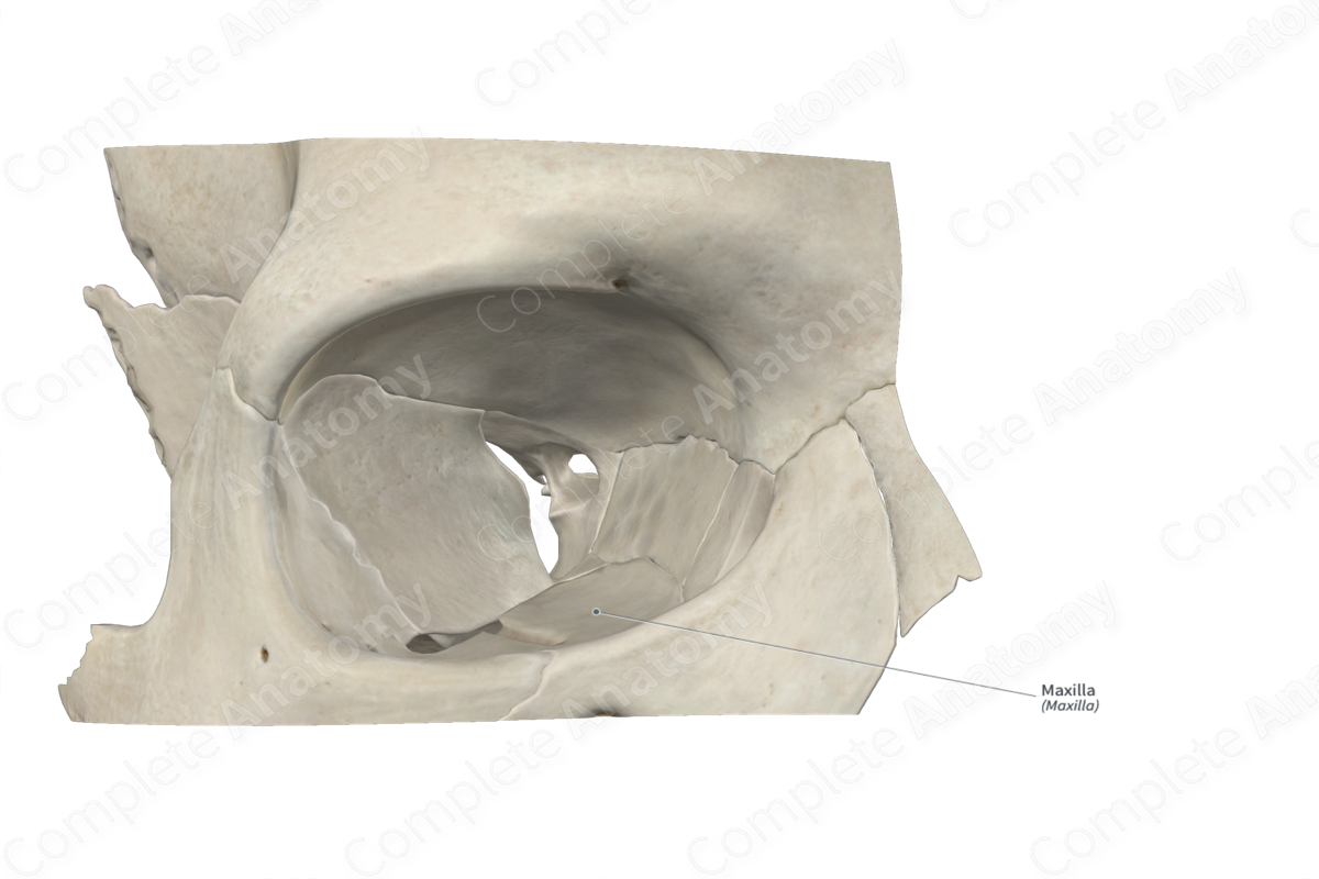
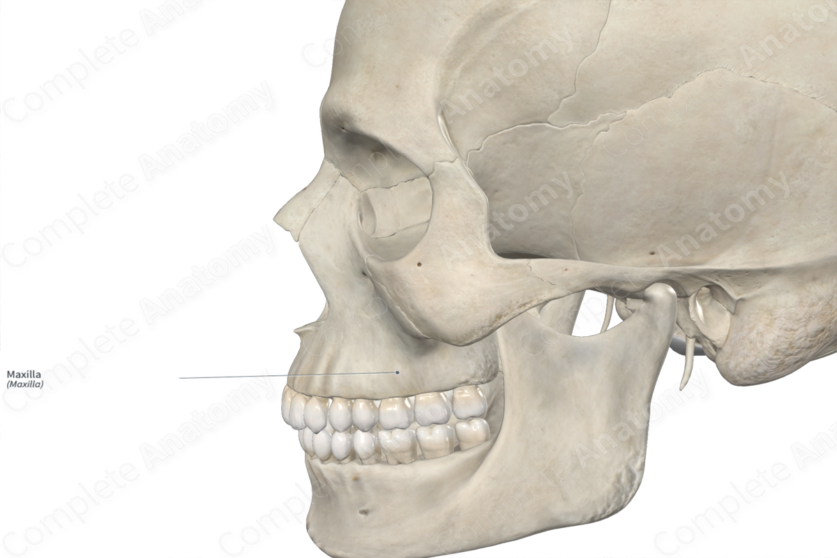
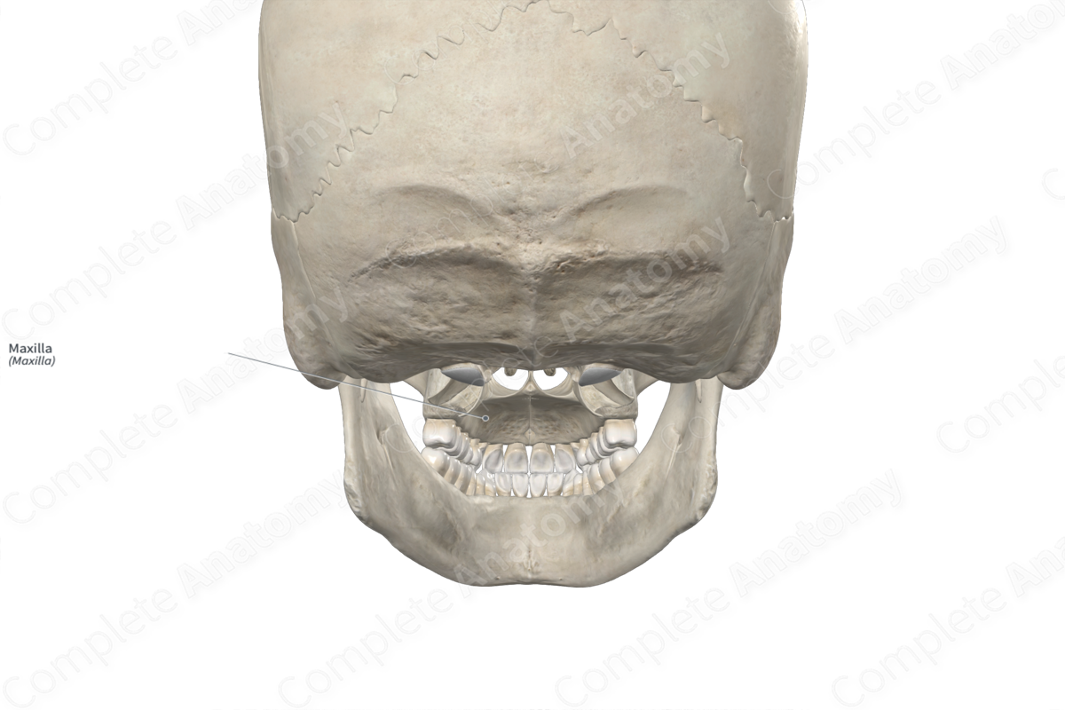
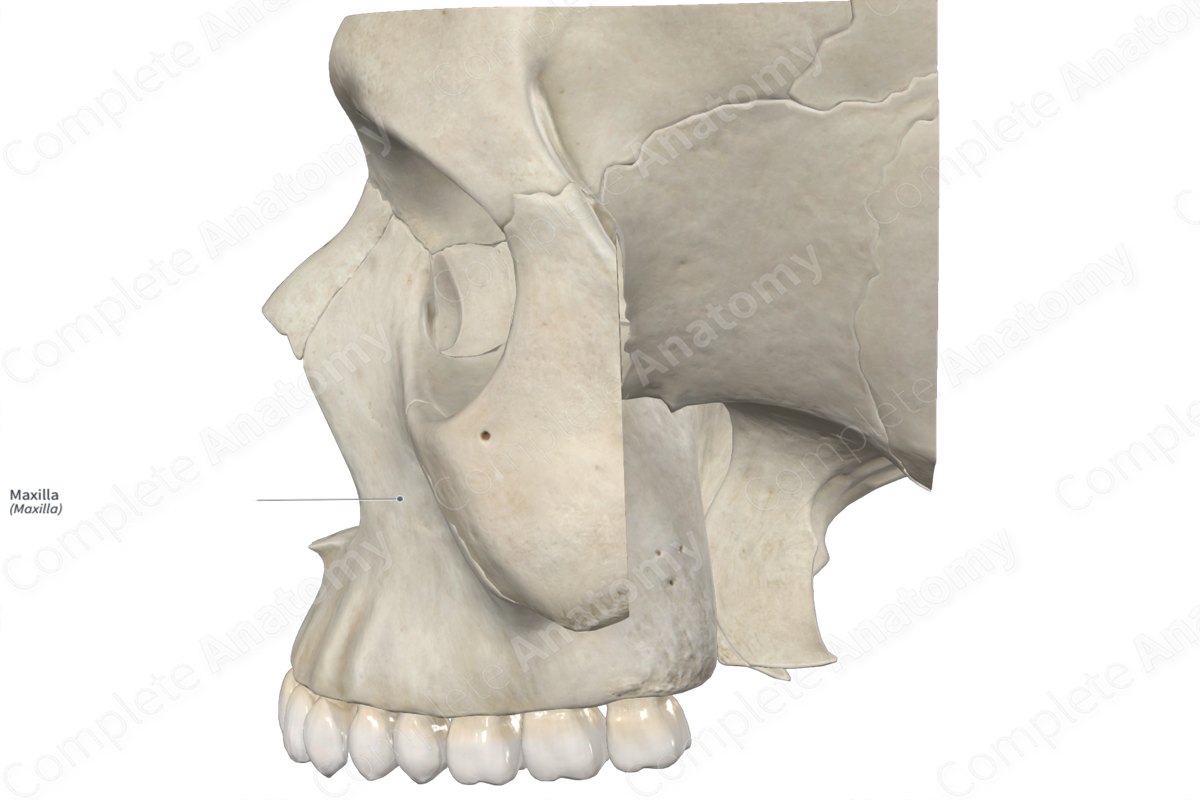
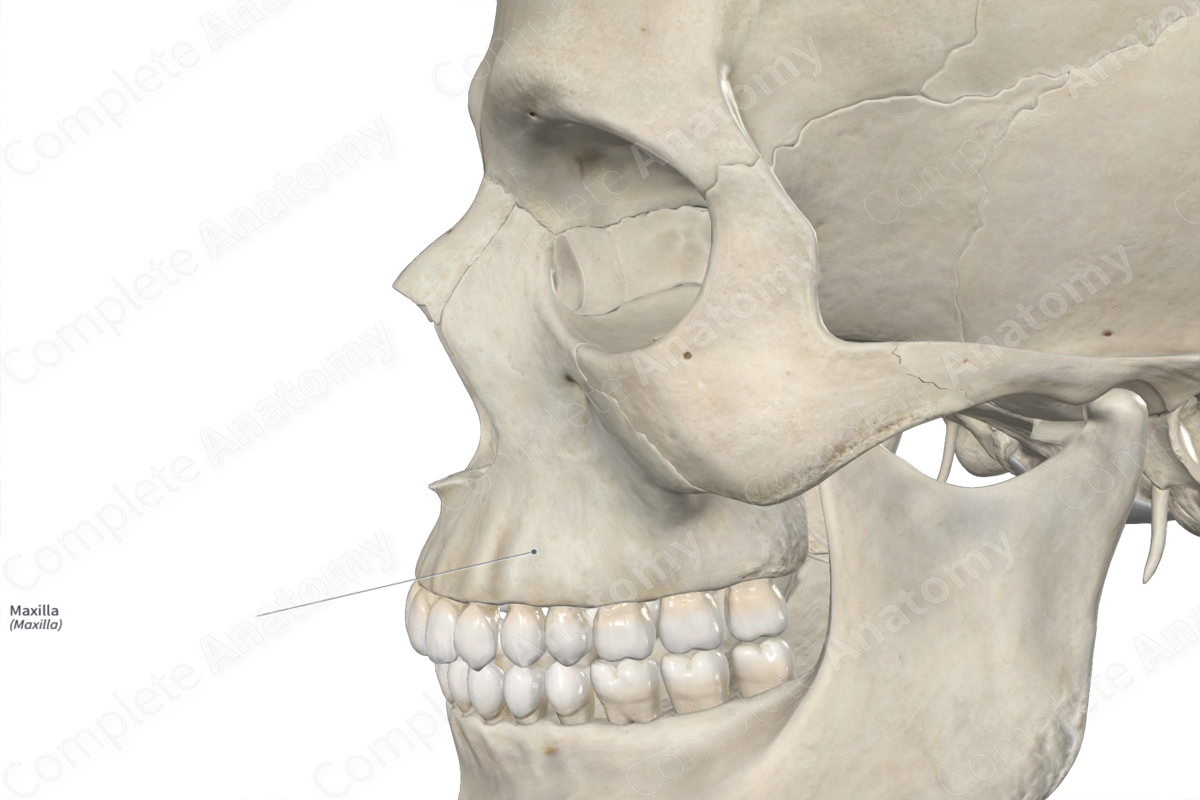
The maxillae (or upper jaw bones) are a pair of large bones found along the anterior aspect of the cranium. They are classified as irregular bones, lodge the maxillary (upper) teeth, and contribute to the formation of the orbits, nasal cavities, bony hard palate, and the viscerocranium. Each maxilla consists of a maxillary sinus and includes the following bony features:
- parts: zygomatic, frontal, alveolar, and palatine processes, and body;
- surfaces: anterior, infratemporal, orbital, nasal, and palatine surfaces, and orbital, ethmoidal, zygomatic, lacrimal, and nasal margins;
- landmarks: canine fossa, canine eminence, maxillary tuberosity, anterior lacrimal crest, and conchal and nasal crests.
More information regarding these bony features can be found in the Parts, Surfaces and Landmarks tabs for this bone.
On its corresponding side, each maxilla is located:
- anterior to the sphenoid, lacrimal and palatine bones;
- superior to the mandible;
- inferior to the frontal and ethmoid bones;
- medial to a zygomatic bone;
- lateral to the vomer, an inferior nasal concha and nasal bone.
Each maxilla articulates with the:
- opposite maxilla at intermaxillary and median palatine sutures;
- lacrimal bone at a lacrimomaxillary suture;
- palatine bone at palatomaxillary and transverse palatine sutures;
- frontal bone at a frontomaxillary suture;
- ethmoid bone at an ethmoidomaxillary suture;
- zygomatic bone at a zygomaticomaxillary suture;
- nasal bone at a nasomaxillary suture;
- vomer;
- inferior nasal concha.
Ossification
Ossification of each maxilla occurs at one ossification center in its body, which appears in utero during the second month and also forms the zygomatic, frontal, alveolar, and palatine processes (Standring, 2016).
Variations
In some individuals:
- accessory infraorbital foramina may be present;
- the maxillary sinus may be underdeveloped or contain a septum;
- the incisive portion of the alveolar process may not be fused with the rest of the maxilla, resulting in the presence of an os incivisum (Tubbs, Shoja and Loukas, 2016).
The maxilla also displays sexual dimorphism, where the maxillae in males are relatively larger than those in females.
Surface Anatomy
Regarding surface anatomy, the body, alveolar process, and palatine process of the maxilla can be palpated.
List of Clinical Correlates
- Fracture of the maxilla (e.g., Le Fort fractures)
- Cleft palate
- Maxillary hypoplasia
References
Gharb, B. B., Rampazzo, A., Kutz, J. E., Bright, L., Doumit, G. and Harter, T. B. (2014) 'Vascularization of the facial bones by the facial artery: implications for full face allotransplantation', Plast Reconstr Surg, 133(5), pp. 1153-65.
Standring, S. (2016) Gray's Anatomy: The Anatomical Basis of Clinical Practice. Gray's Anatomy Series 41st edn.: Elsevier Limited.
Tubbs, R. S., Shoja, M. M. and Loukas, M. (2016) Bergman's Comprehensive Encyclopedia of Human Anatomic Variation. Wiley.
Learn more about this topic from other Elsevier products

