Quick Facts
Origin: Medulla oblongata of the brainstem.
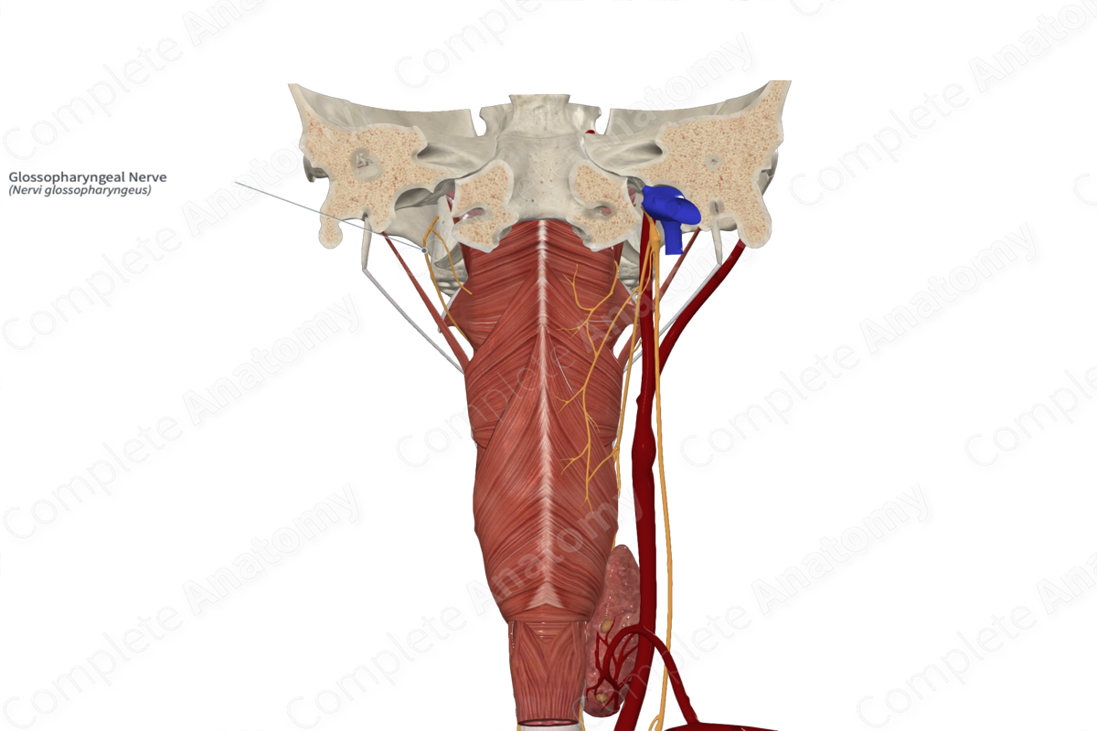
Course: Runs laterally from the brainstem to the jugular foramen. Emerges below the skull posterior to the internal jugular vein and internal carotid artery. Passes between these vessels, moving anteriorly and inferiorly to wrap around the stylopharyngeus muscle before terminating into branches between the superior and middle pharyngeal constrictor muscle.
Branches: Tympanic, carotid sinus, pharyngeal, stylopharyngeal, tonsillar, and lingual branches. It contributes to the tympanic plexus, lesser petrosal nerve, pharyngeal plexus, and several variable communications to other cranial nerves.
Supply: Parasympathetic: parotid gland; Motor: stylopharyngeus muscle; Sensory: tympanic cavity, auditory tube, oropharynx, the external ear, posterior one third of the tongue (both general sense and taste), carotid body, and carotid sinus.
Related parts of the anatomy
Origin
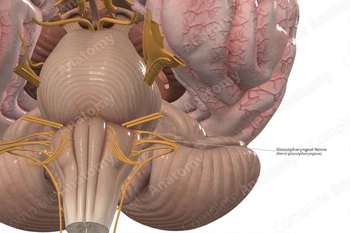
The glossopharyngeal nerve originates in the medulla oblongata of the brainstem. The specific brainstem nuclei giving rise to the glossopharyngeal nerve include the nucleus ambiguous, nucleus of the solitary tract, inferior salivatory nucleus, and the spinal nucleus of trigeminal nerve (Norton, 2016; Shoja & Oyesiku, 2014).
Course
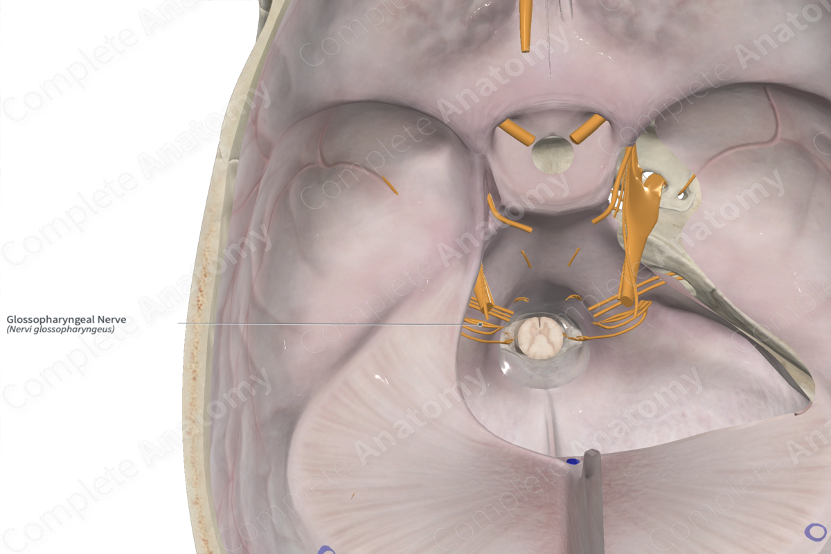
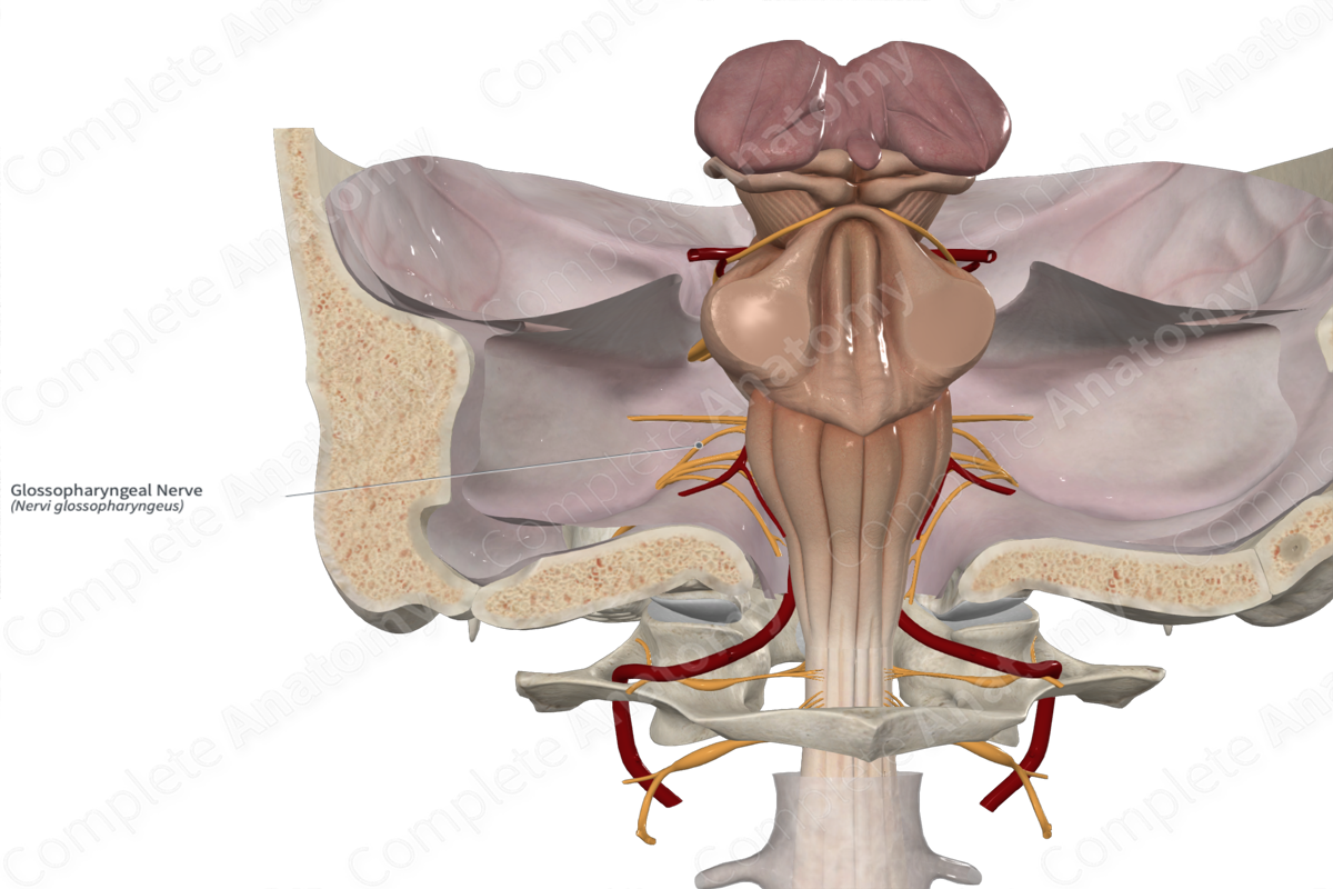
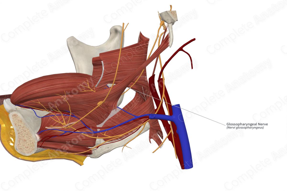
The glossopharyngeal nerve exits the medulla laterally, just posterior to the pontomedullary junction. From here it runs laterally to and through the jugular foramen to exit the skull. After exiting the skull, the glossopharyngeal nerve is located medial to the styloid process. It runs down and forward, passing between the internal jugular vein and the internal carotid artery, and along the posterior border of stylopharyngeus muscle. It wraps around stylopharyngeus muscle to the lateral and anterior surface, before passing into the space between the superior and middle pharyngeal constrictor muscles. As it passes through this space and deep to hyoglossus muscle. The glossopharyngeal nerve terminates in its final branches (Norton, 2016; Shoja & Oyesiku, 2014).
Branches
The glossopharyngeal nerve has branches that split off at many points.
The tympanic nerve splits off at the level of the inferior ganglion. It heads superiorly into the tympanic cavity where it contributes to the tympanic plexus and lesser petrosal nerve.
The carotid sinus nerve branches off at or just below the inferior ganglion. It runs down to the carotid sinus and carotid body.
The stylopharyngeal branch is given off as the glossopharyngeal nerve travels down the stylopharyngeus muscle.
The pharyngeal branches are given off as the glossopharyngeal nerve wraps around stylopharyngeus muscle. They travel inferiorly to the posterior surface of the middle pharyngeal constrictor where they mix with vagal and sympathetic fibers to form the pharyngeal plexus.
The tonsillar branches split off when the glossopharyngeal nerve is located roughly between the superior and middle pharyngeal constrictor muscles. They ascend to the palatine tonsils.
The lingual branches are the last of the terminal branches of the glossopharyngeal nerve. Deep to hyoglossus, they move medially to the posterior one third of the tongue.
There are a number of variable communications between the glossopharyngeal nerve and other cranial nerves found in proximity in the neck. The best characterized communication is that between the vagus nerve and the glossopharyngeal nerve. Fibers whose cell bodies are in the superior glossopharyngeal ganglion that carries sensation from portions of the external ear travel back via the vagus nerve before passing to the glossopharyngeal nerve.
Supplied Structures
The glossopharyngeal nerve is a mixed nerve conveying parasympathetic, motor, and sensory innervation.
Parasympathetic efferent fibers from the tympanic nerve target the parotid gland via the lesser petrosal nerve.
Branchial motor efferent fibers from the stylopharyngeal branch innervate the stylopharyngeus muscle.
General somatic afferent fibers convey sensation from the posterior one third of the tongue as well as a small portion of the skin of the external ear. These fibers travel in the lingual branch and the communication with the vagus nerve, respectively.
General visceral afferent fibers convey sensation from the mucosal lining of the tympanic cavity, auditory tube, oropharynx, carotid body, and carotid sinus. These are carried by the tympanic nerve, the pharyngeal branches, and the carotid sinus nerve, respectively.
Special visceral afferent fibers carry taste sensation from the posterior one third of the tongue via the lingual branches.
List of Clinical Correlates
—Gag reflex
References
Norton, N. S. (2016) Netter's Head and Neck Anatomy for Dentistry E-Book. Elsevier Health Sciences.
Shoja, M. M. & Oyesiku, N. M. (2014) Clinical anatomy of the cranial nerves. Clin Anat, 27(1), 2-3.
Learn more about this topic from other Elsevier products

Anatomy of the glossopharyngeal nerve (CN IX)

Master Anatomy of the glossopharyngeal nerve (CN IX): structure, function, and composition. High-yield review with labeled diagrams and practice quizzes.
