Unfortunately we don't fully support your browser. If you have the option to, please upgrade to a newer version or use Mozilla Firefox, Microsoft Edge, Google Chrome, or Safari 14 or newer. If you are unable to, and need support, please send us your feedback.

Elsevier
Publish with us
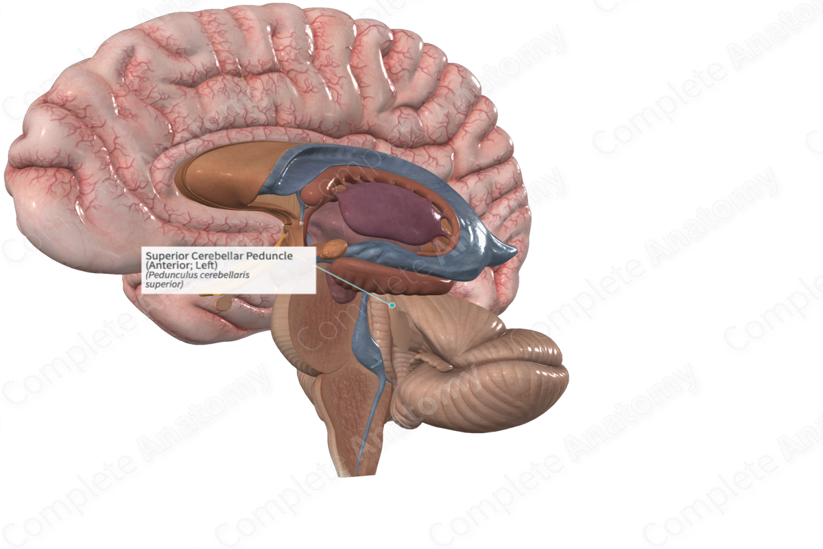
Superior Cerebellar Peduncle (Anterior; Left)
Nervous System

Superior Cerebellar Peduncle (Anterior; Left)

Pedunculus cerebellaris superior

Read more

Quick Facts

The superior cerebellar peduncle (aka rostral cerebellar peduncle, or brachium conjunctivum) consists of a large bundle of nerve fibers emerges from the cerebellum along the lateral wall of the fourth ventricle into the tegmentum where most of its fibers decussate.

The fibers originate from the dentate and interpositus nuclei. They terminate in the contralateral red nucleus and parts of the ventrolateral, ventral intermediate and central lateral nuclei of the thalamus.

Complete Anatomy
The world's most advanced 3D anatomy platform
Try it for Free

Learn more about this topic from other Elsevier products

Superior Cerebellar Peduncle

ScienceDirect image

The superior cerebellar peduncle is the principal efferent pathway of axons leaving the globose, emboliform, and dentate nuclei (the intermediate and lateral nuclei), and also carries afferents into the cerebellum from the ventral spinocerebellar tract, the tectocerebellar tract, the rubrocerebellar tract, and the noradrenergic projections from the locus coeruleus.

Explore on ScienceDirectopens in new tab/window

Complete Anatomy

The world's most advanced 3D anatomy platform

Complete Anatomy