Structure
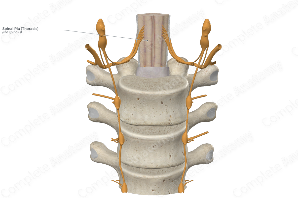
Spinal pia mater is similar to cranial pia mater in both structure and position. The spinal pia mater is wrapped directly on the spinal cord and invests within the ventral median fissure and dorsal sulci of the spinal cord. Spinal pia mater is a very thin, largely transparent, fibrous membrane.


Related parts of the anatomy
Key Features/Anatomical Relations
Spinal pia mater adheres directly to the spinal cord and invests within the ventral median fissure and the dorsal sulci of the spinal cord. It is only free from the spinal cord as it extends laterally to form denticulate ligaments, and inferiorly, as it forms the filum terminale below the inferior terminus of the spinal cord. Because it is so thin and largely transparent, it is generally not visible. It appears somewhat opaque, with a white to silver color, as it forms the denticulate ligaments and filum terminale.
Learn more about this topic from other Elsevier products

