
Unfortunately we don't fully support your browser. If you have the option to, please upgrade to a newer version or use Mozilla Firefox, Microsoft Edge, Google Chrome, or Safari 14 or newer. If you are unable to, and need support, please send us your feedback.

Elsevier
Publish with us
Denticulate Ligament (Thoracic; Left)
Nervous System

Denticulate Ligament (Thoracic; Left)

Ligamentum denticulatum

Read more

Structure

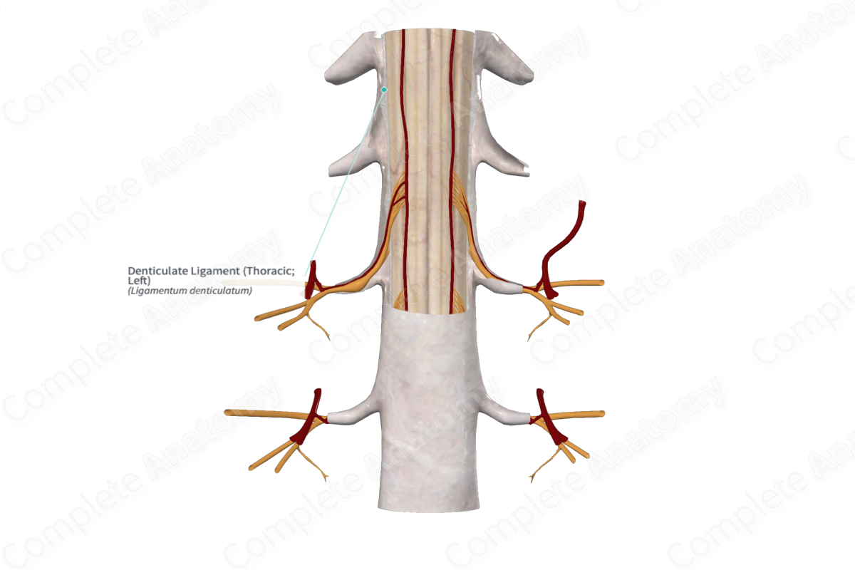
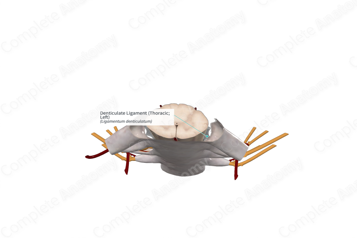
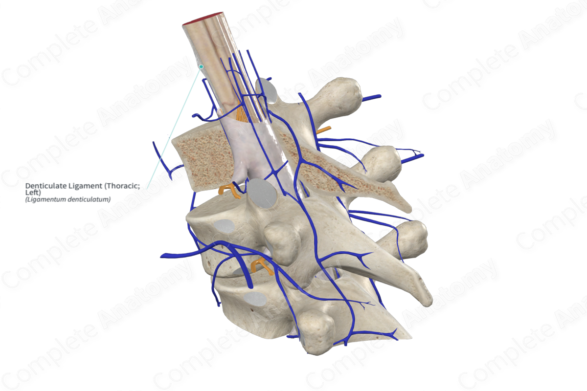
The denticulate ligaments are thin sheets of collagen that extend laterally from the spinal pia mater. They extend out and form triangular shaped attachments to the overlying dura mater.

Complete Anatomy
The world's most advanced 3D anatomy platform
Try it for Free

Key Features/Anatomical Relations

The denticulate ligaments extend from the foramen magnum rostrally down to the level of the T12 vertebra caudally. On each side, there are typically 18-21 triangular extensions which attach to the spinal dura mater, lateral to the spinal cord. They are thought to help stabilize the spinal cord. The lateral extensions of the denticulate ligaments can be seen laying between the dorsal and ventral nerve roots.

Learn more about this topic from other Elsevier products

Denticulate Ligaments

ScienceDirect image

The denticulate ligaments are fibrous structures that help to anchor the spinal cord in place.

Explore on ScienceDirect(opens in new tab/window)

Complete Anatomy

The world's most advanced 3D anatomy platform

Complete Anatomy