
Structure
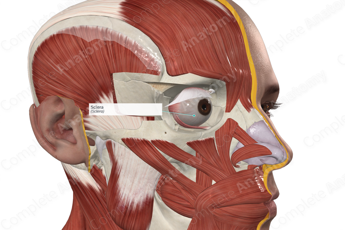
The sclera forms part of the tough white outer fibrous layer for most of the eyeball, covering more than 93% of the surface of the eyeball. Only a small portion of the sclera is visible when the eyeball sits in situ in the orbital cavity, since the sclera is replaced by the transparent cornea in the anterior portion of the eyeball (Standring, 2016).
The sclera can vary in thickness, ranging from about 0.3-1mm. It is composed of collagen fibers that are interlaced, providing the strength and flexibility of the eyeball. It is the organization of these fibers that lends the sclera its opaque appearance.


Key Features & Anatomical Relations
The sclera is continuous anteriorly with the cornea at the corneoscleral junction and is attached to the ciliary body. Additionally, it is covered anteriorly by the conjunctiva, which reflects onto it from the posterior surface of the eyelids.
Posteriorly, the sclera is pierced by the optic nerve medial and slightly inferior to the midline of the eyeball. The outer surface of the sclera is continuous with the dura mater that covers the optic nerve, while the inner surface forms a plate with many perforations, the lamina cribrosa sclerae. Fibers from the optic nerve perforate through these small perforations, along with the central retinal artery and vein.
The internal surface of the sclera is lined by the choroid, thus, separating the sclera from the retina.
Function
The fibrous layer of the eyeball provides the overall shape of the eyeball, while withstanding intraocular pressure. The tough fibrous sclera protects the eye from damage from the external environment. Additionally, the sclera provides the attachment for extraocular (extrinsic) muscles of the eyeball.
List of Clinical Correlates
- Scleritis
- Jaundice
References
Standring, S. (2016) Gray's Anatomy: The Anatomical Basis of Clinical Practice. Gray's Anatomy Series: Elsevier Limited.
Learn more about this topic from other Elsevier products

