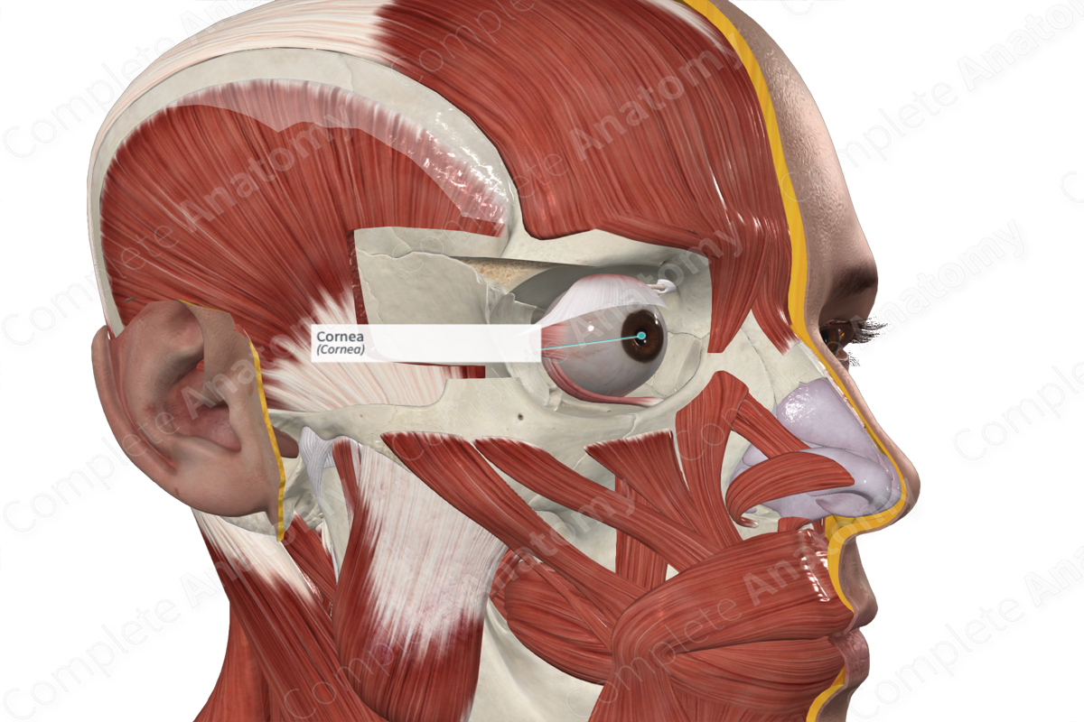
Structure
The cornea forms a small part of the tough outer fibrous layer of the eyeball, covering about 7% of the external surface. The sclera forms the remainder of the fibrous outer layer of the eyeball.
The cornea is convex anteriorly, projecting as a dome from the sclera and varies in thickness, from about 670μm at the corneoscleral junction (where the cornea and sclera become continuous) to 520μm at the center of the cornea. The external surface of the cornea is covered by a tear film, producing lubrication and hydration to its surface.
The cornea consists of 5 layers including, from anterior to posterior, the corneal epithelium, anterior limiting lamina, stroma, posterior limiting layer, and endothelium.
The corneal epithelium makes up 10% of the corneal thickness. It is composed of 5-6 layers of cells that aid in protecting the surface of the eye from abrasion, preventing the entry of pathogens into the eyeball, and allowing the passage of small molecules, water, and ions.
Directly behind the corneal epithelium lies the anterior limiting lamina or Bowman’s layer. It is a thin layer composed of dense collagen fibrils. The thickest layer of the cornea is the substantia propria, or stroma. It is composed of 200-250 layers of parallel collagen fibrils with fibroblasts sitting in between.
The posterior limiting lamina, or Descemet’s layer lines the stroma posteriorly and acts as a basement membrane from the underlying endothelium. The endothelium is a single cell layer of squamous cells lining the posterior aspect of the cornea (Standring, 2016).


Key Features & Anatomical Relations
The cornea is continuous with the sclera at the limbus. Here, the epithelial layer of the cornea merges with the epithelium of the conjunctiva. The anterior limiting lamina terminates, and the corneal stroma loses its collagen density. The posterior limiting lamina becomes continuous with the trabecular meshwork, while the endothelium of the cornea is continuous with the epithelium covering the trabeculae.
Function
The cornea is the most powerful contributor to the reflective power of the eye, accounting for 70% of the refractive power. Therefore, changes to the shape of the cornea will have significant implications for vision.
List of Clinical Correlates
- Astigmatism
References
Standring, S. (2016) Gray's Anatomy: The Anatomical Basis of Clinical Practice. Gray's Anatomy Series: Elsevier Limited.
Learn more about this topic from other Elsevier products

