Quick Facts
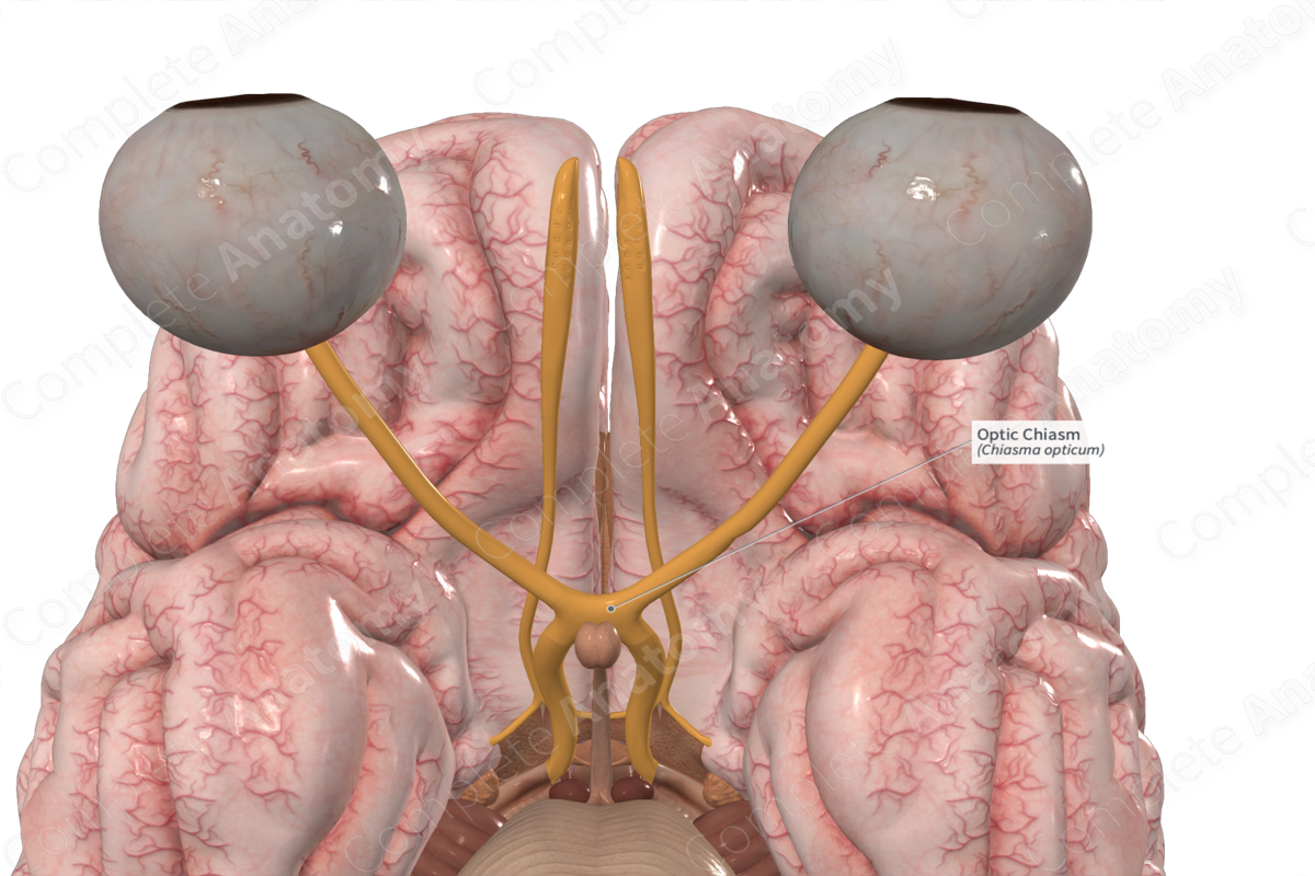
Origin: Formed by the merging of fibers from the optic nerves.
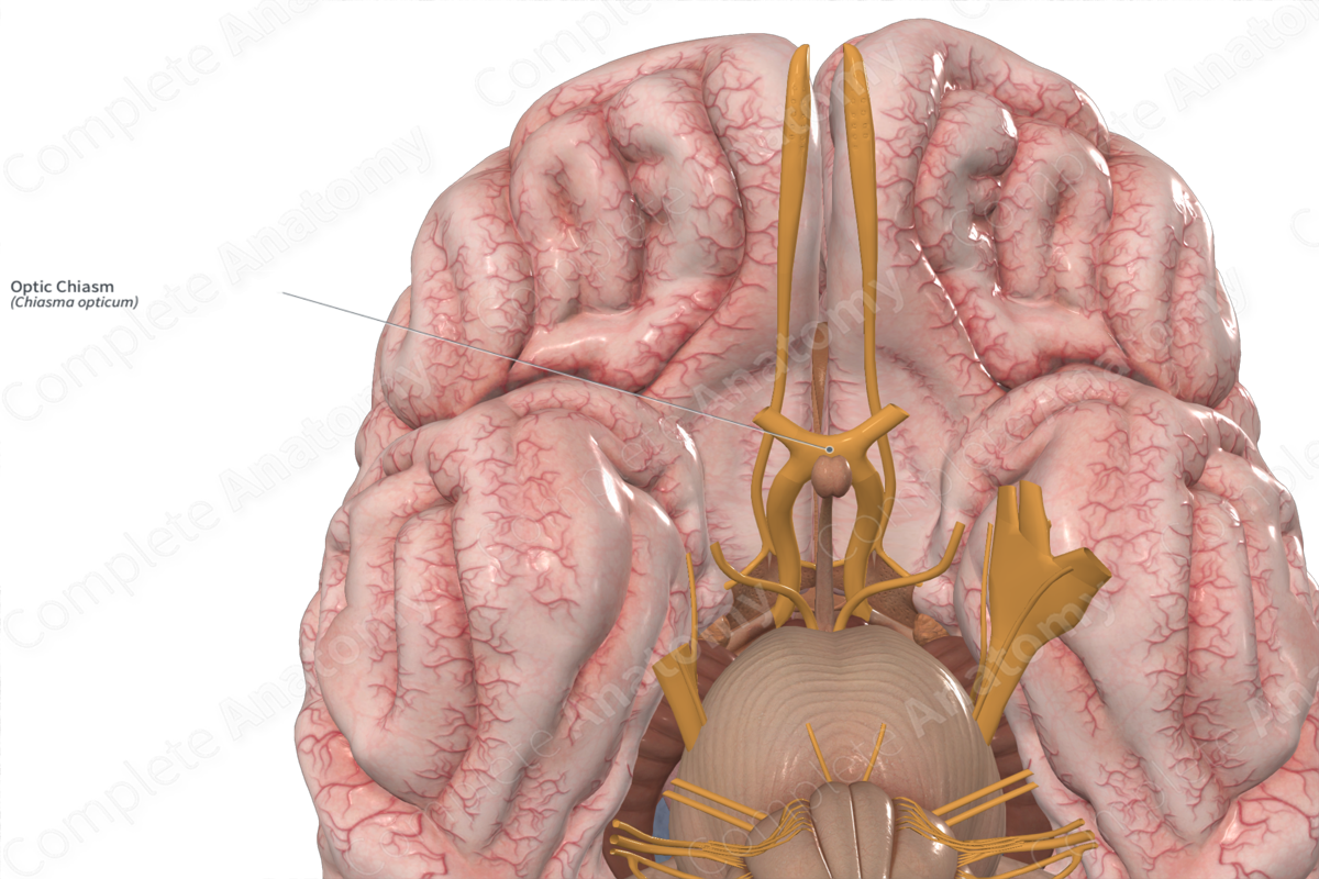
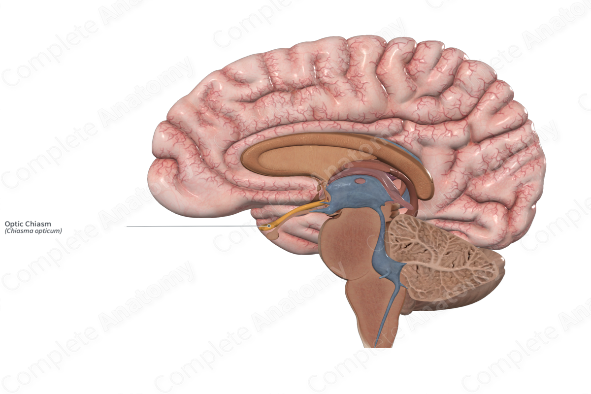
Location: Sits superior and anterior to the pituitary gland on the midline.
Branches: Optic tracts.
Supply: Conveys visual information towards the cortex.

Related parts of the anatomy
Location
The optic chiasm occupies a space inferior to the ventral midline of the brain, just anterior and superior to the location of the pituitary gland. It represents the crossing of nasal retinal fibers from one optic nerve to the contralateral optic tract.
Branches
The optic chiasm splits into the two optic tracts.
Supplied Structures
The optic chiasm is sensory and conveys visual information towards the cortex. It sends axons into the optic tracts.
By allowing nasal retinal fibers from one optic nerve to cross to the contralateral optic tract, the optic chiasm allows all right visual field information to travel to the left hemisphere of the brain, and all left visual field information to travel to the right hemisphere.
List of Clinical Correlates
- Visual deficits
Learn more about this topic from other Elsevier products

