Quick Facts
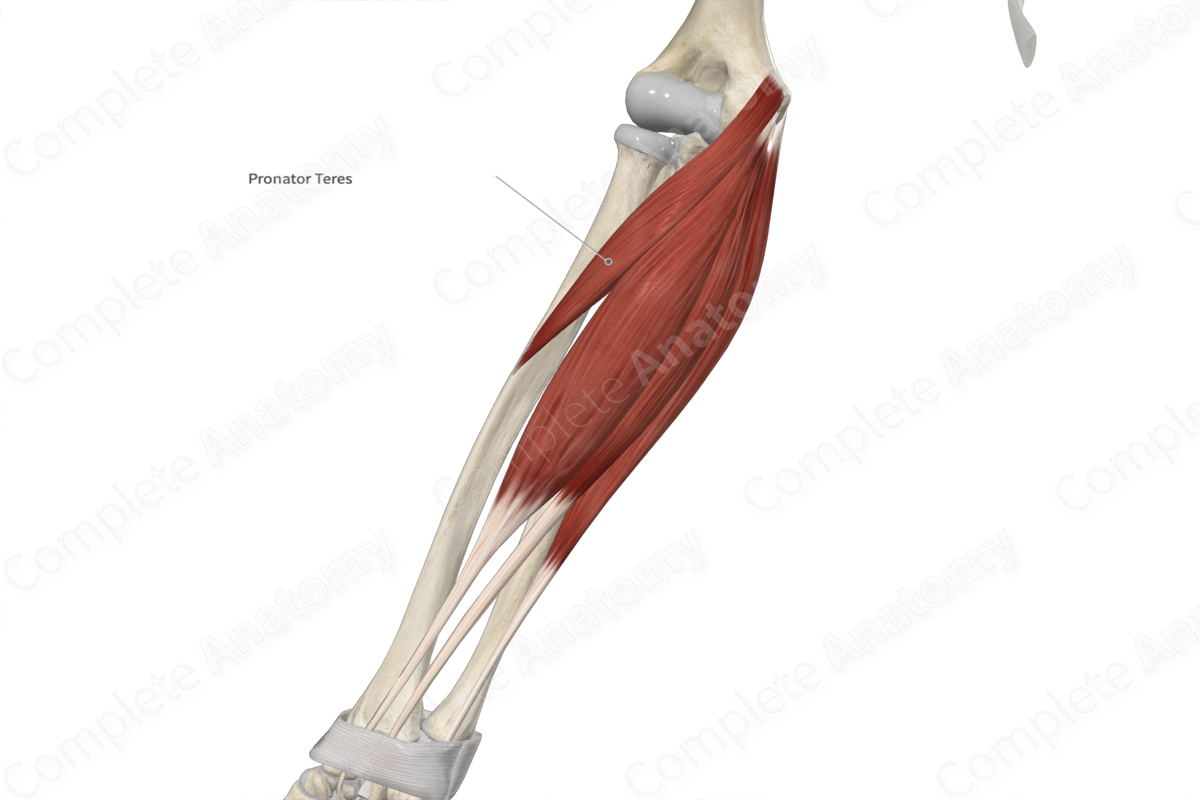
Origin: Medial supracondylar ridge of humerus, via the common flexor tendon, and medial aspect of coronoid process of ulna.
Insertion: Middle one third of anterolateral aspect of radius.
Action: Pronates forearm at radioulnar joints.
Innervation: Median nerve (C6-C7).
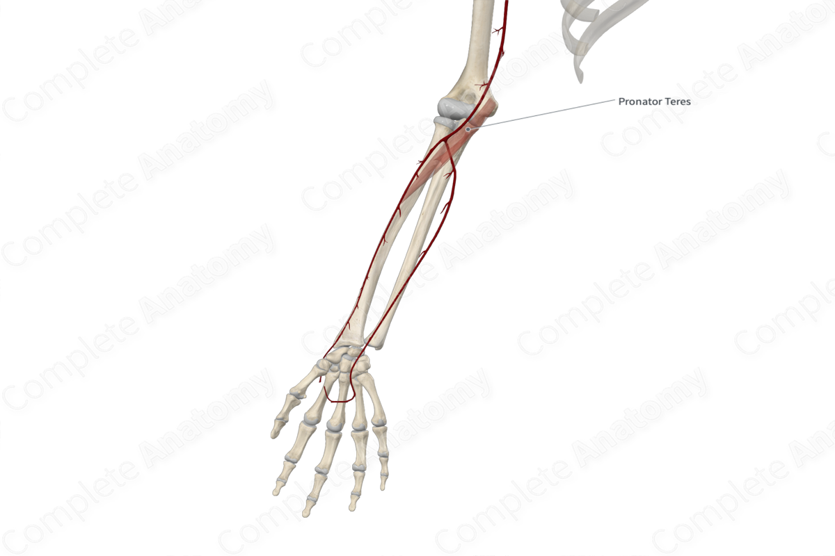
Arterial Supply: Anterior branch of ulnar recurrent artery, inferior ulnar collateral, common interosseous, ulnar, and radial arteries.
Related parts of the anatomy
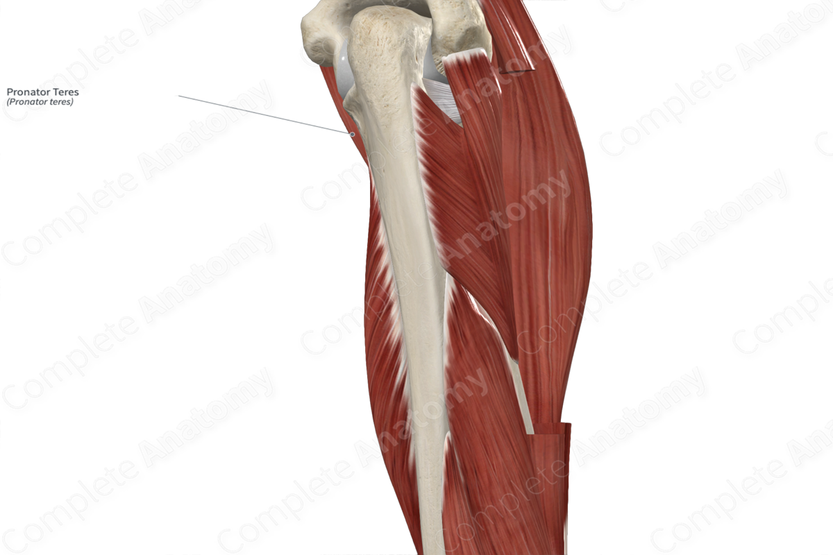
Origin
The pronator teres muscle consists of two heads:
- a large humeral head, which originates, via the common flexor tendon, from the medial supracondylar ridge of the humerus and adjacent antebrachial fascia;
- a small ulnar head, which lies deep to the humeral head and originates from the medial aspect of the coronoid process of ulna.
The common flexor tendon is the combined tendon for all five superficial muscles in the anterior compartment of the forearm, which are the:
- pronator teres muscle;
- flexor carpi radialis muscle;
- palmaris longus muscle;
- flexor carpi ulnaris muscle;
- flexor digitorum superficialis muscle.
Insertion
The fibers of the pronator teres travel inferolaterally to the middle of the forearm and insert, via a flat tendon, onto the middle one third of the anterolateral aspect of radius.
Key Features & Anatomical Relations
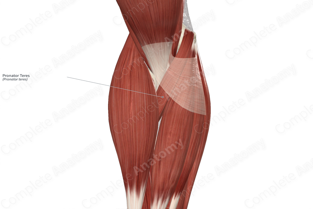
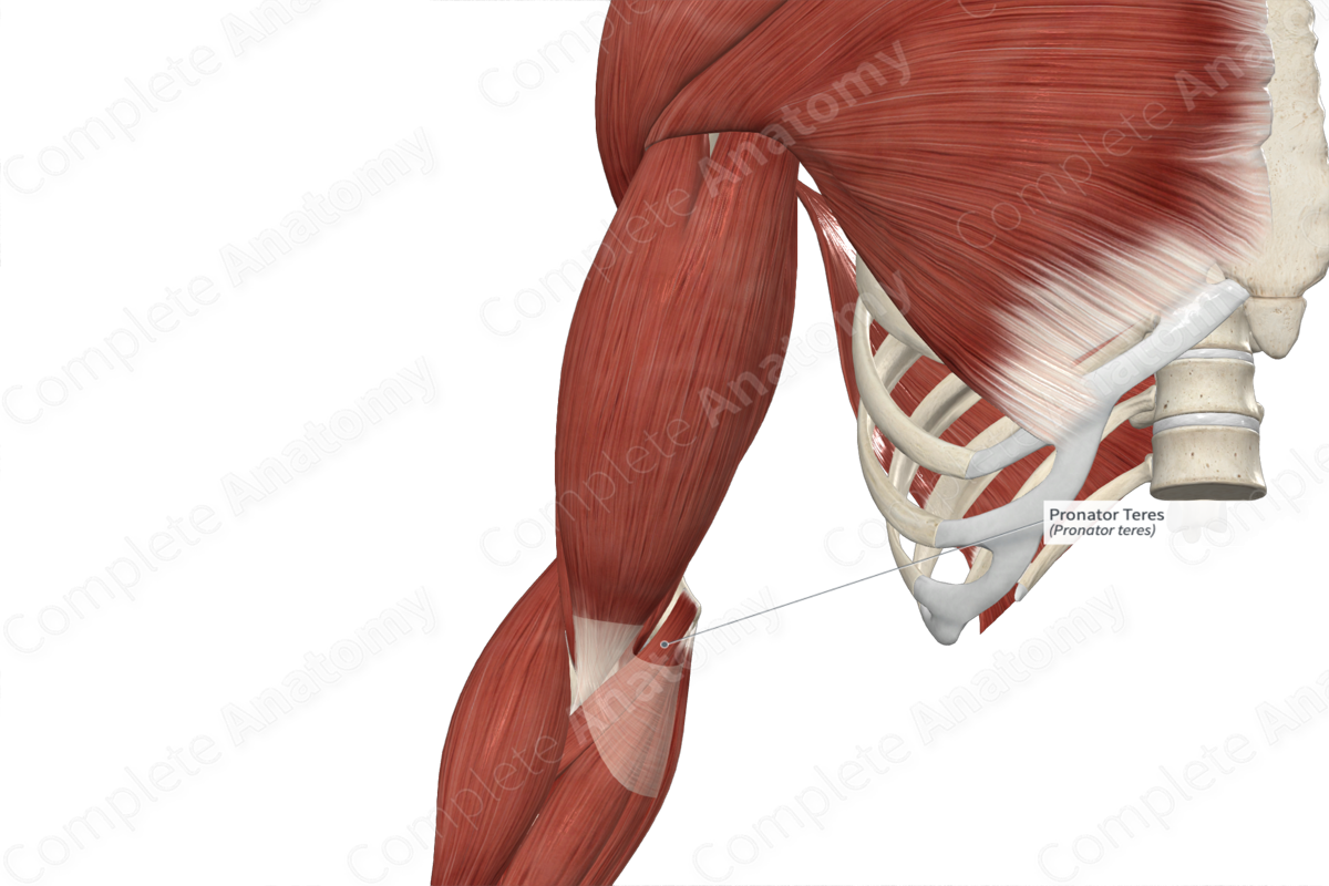
The pronator muscle is one of the muscles of the superficial part of anterior compartment of the forearm. It is a fusiform type of skeletal muscle. It is located:
- anterior (superficial) to the flexor digitorum superficialis muscle and ulnar artery;
- posterior (deep) to the brachioradialis muscle (at its distal end);
- lateral to the flexor carpi radialis muscle.
In order to enter the anterior compartment of the forearm, the median nerve travels between the humeral and ulnar heads of the pronator teres muscle. Furthermore, the lateral margin of the pronator teres muscle defines the medial boundary of the cubital fossa.
Actions & Testing
The pronator teres muscle is involved in multiple actions:
- pronates the forearm at the radioulnar joints;
- assists in flexion of the forearm at the elbow joint.
The pronator teres muscle can be tested by pronating the forearm against resistance, during which it can be palpated (Standring, 2016).
List of Clinical Correlates
- Medial epicondylitis (golfer’s elbow)
References
Standring, S. (2016) Gray's Anatomy: The Anatomical Basis of Clinical Practice. Gray's Anatomy Series 41st edn.: Elsevier Limited.
Learn more about this topic from other Elsevier products

