Quick Facts
Origin: Medial epicondyle of humerus, via the common flexor tendon, sublime tubercle of ulna, and proximal half of anterior border of radius.
Insertion: Palmar aspects of the bodies of middle phalanges of the index, middle, ring, and little fingers.
Action: Flexes index, middle, ring, and little fingers.
Innervation: Median nerve (C7-T1).
Arterial Supply: Anterior branch of ulnar recurrent artery, ulnar and radial arteries.


Related parts of the anatomy
Origin
The flexor digitorum superficialis muscle consists of two heads:
- a humeroulnar head, which originates from the medial epicondyle of the humerus via the common flexor tendon, the sublime tubercle of ulna, and the ulnar collateral ligament of the elbow joint;
- a radial head, which originates from the proximal half of the anterior border of the body of the radius.
The common flexor tendon is the combined tendon for all five superficial muscles in the anterior compartment of the forearm, which are the:
- pronator teres muscle;
- flexor carpi radialis muscle;
- palmaris longus muscle;
- flexor carpi ulnaris muscle;
- flexor digitorum superficialis muscle.
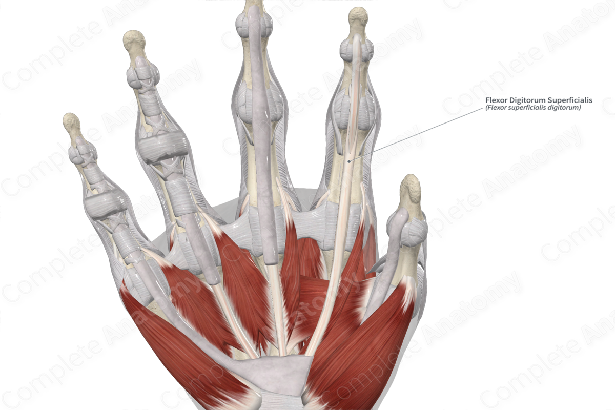
Insertion
The fibers of the flexor digitorum superficialis muscle travel inferiorly to the hand and insert onto the palmar aspects of the bodies of middle phalanges of the index, middle, ring, and little fingers.
Key Features & Anatomical Relations
The flexor digitorum superficialis muscle is one of the muscles of the superficial part of anterior compartment of the forearm. It is a long, broad, fusiform type of skeletal muscle that divides into four tendons. The tendons for the middle and ring fingers lie superficial to the tendons for the index and ring fingers. In the forearm and hand, each of these tendons travels superficial to the tendons of the flexor digitorum profundus muscle.
At the wrist, the tendons of both muscles travel together, deep to the flexor retinaculum of hand, where they pass through the common flexor sheath. The tendons of both muscles travel through the fibrous sheaths that are found along the palmar aspect of each finger. Along each proximal phalanx, the tendons of the flexor digitorum profundus pass through the tendons of the flexor digitorum superficialis and travel to their insertion sites.
The flexor digitorum superficialis muscle is located:
- anterior (superficial) to the flexor digitorum profundus and flexor pollicis longus muscles, and the median nerve;
- posterior (deep) to the flexor carpi radialis, palmaris longus, and flexor carpi ulnaris muscles;
- medial to the brachioradialis muscle.
In order to enter the anterior compartment of the forearm, the median nerve and ulnar artery travel between the humeroulnar and radial heads of the flexor digitorum superficialis muscle.
Actions & Testing
The flexor digitorum superficialis muscle is involved in multiple actions:
- flexes the middle phalanges at the proximal interphalangeal joints of the index, middle, ring, and little fingers;
- assists in flexion of the proximal phalanges at the metacarpophalangeal joints of the same fingers;
- assists in flexion of the hand at the radiocarpal (wrist) joint.
This muscle is capable of flexing each finger independently of the other fingers.
The flexor digitorum superficialis muscle can be tested by flexing the middle phalanx at the proximal interphalangeal joint while the other three fingers are held in full extension. This extended position of the other three fingers prevents the flexor digitorum profundus muscle from contracting (Standring, 2016).
List of Clinical Correlates
- Medial epicondylitis (golfer’s elbow)
References
Standring, S. (2016) Gray's Anatomy: The Anatomical Basis of Clinical Practice. Gray's Anatomy Series 41st edn.: Elsevier Limited.
Learn more about this topic from other Elsevier products

