Quick Facts
Origin: Medial epicondyle of humerus, via common flexor tendon, proximal two thirds of body of ulna, and olecranon of ulna.
Insertion: Pisiform, hook of hamate, and palmar aspect of base of fifth metacarpal bone.
Action: Flexes and adducts hand at radiocarpal (wrist) joint.
Innervation: Ulnar nerve (C7-T1).
Arterial Supply: Ulnar artery and posterior branch of ulnar recurrent artery.
Related parts of the anatomy
Origin
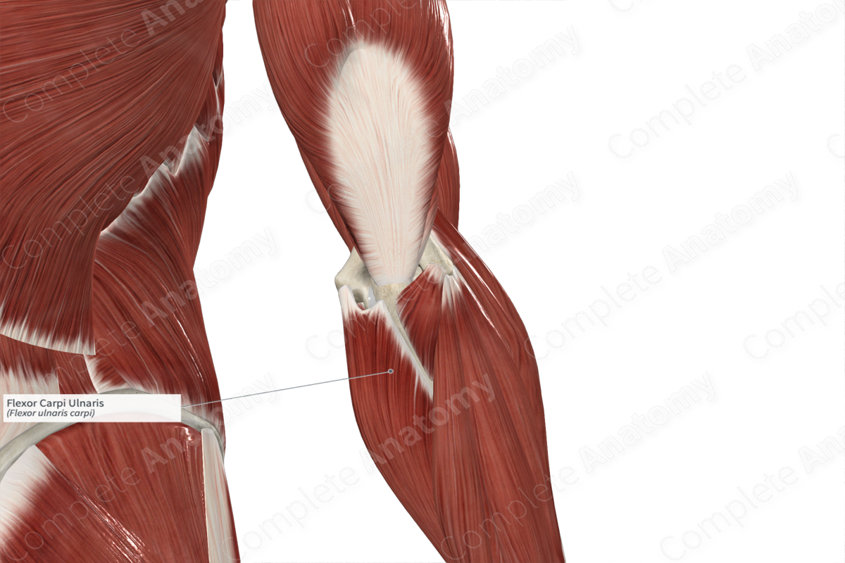
The flexor carpi ulnaris muscle consists of two heads:
- a small humeral head, which originates, via the common flexor tendon, from the medial epicondyle of the humerus;
- a large ulnar head, which originates from the posteromedial aspects of both the proximal two thirds of the body of ulna and the olecranon.
The common flexor tendon is the combined tendon for all five superficial muscles in the anterior compartment of the forearm, which are the:
- pronator teres muscle;
- flexor carpi radialis muscle;
- palmaris longus muscle;
- flexor carpi ulnaris muscle;
- flexor digitorum superficialis muscle.
Insertion
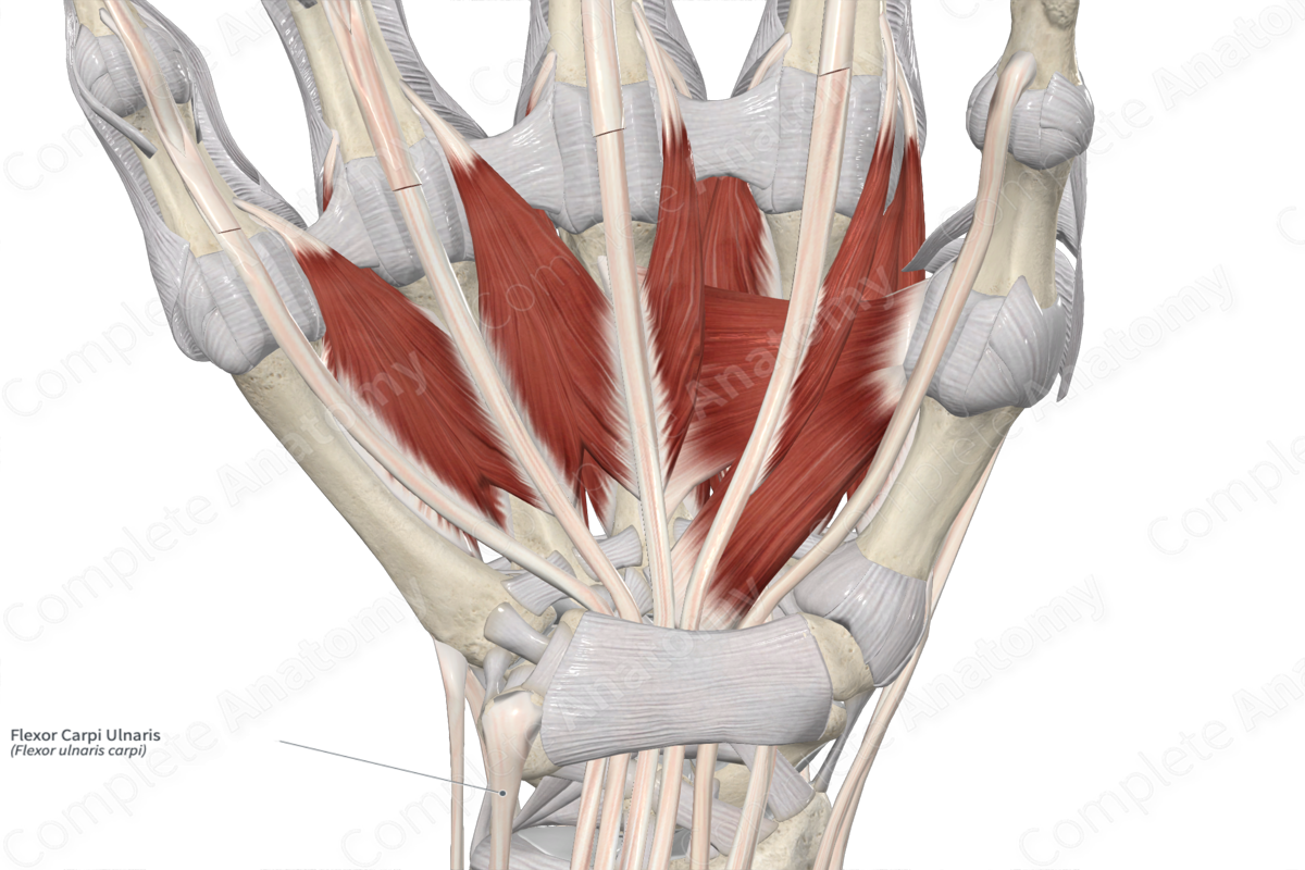
The fibers of the flexor carpi ulnaris muscle travel inferiorly to the hand and insert, via a long tendon, onto the pisiform bone. This insertion site is further extended from the pisiform bone to the:
- palmar aspect of the hook of hamate bone, via the pisohamate ligament;
- palmar aspect of the base of fifth metacarpal bone, via the pisometacarpal ligament.
Key Features & Anatomical Relations
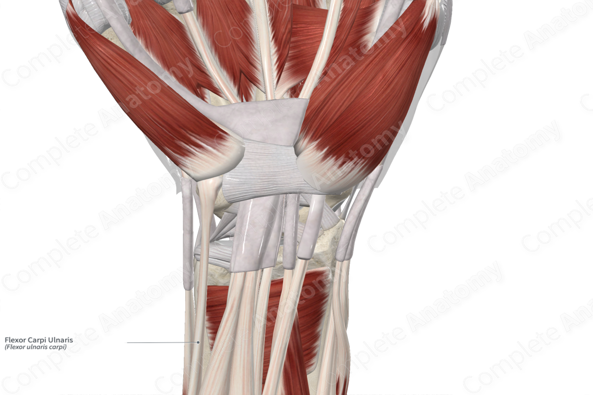
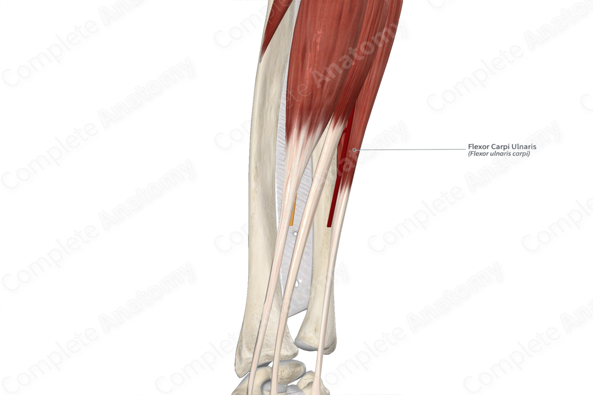
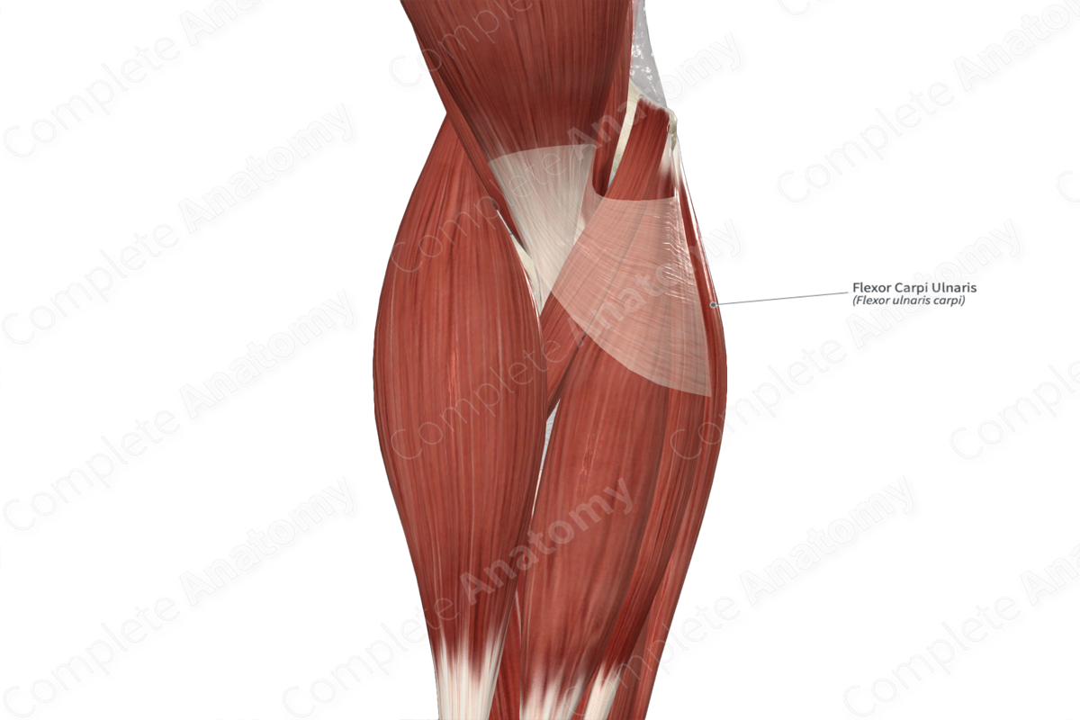
The flexor carpi ulnaris muscle is one of the muscles of the superficial part of anterior compartment of the forearm. At the distal one third of the forearm, the muscle belly gives rise to a long tendon, which travels inferomedially. At the wrist, the tendon travels deep to the palmar carpal ligament to its insertion sites.
The flexor carpi ulnaris muscle is located:
- anterior to the extensor carpi ulnaris muscle;
- superficial to the ulnar artery and nerve;
- medial to all the other superficial muscles in the anterior compartment of the forearm (at its muscle belly), and the ulnar artery and ulnar nerve (at its distal tendon).
In order to enter the anterior compartment of the forearm, the ulnar nerve travels deep to the tendinous arch that is formed between the humeral and ulnar heads of the flexor carpi ulnaris muscle.
Actions & Testing
The flexor carpi ulnaris muscle is involved in multiple actions:
- flexes the hand at the radiocarpal (wrist) joint, which occurs when the flexor carpi radialis muscle contracts simultaneously with it;
- adducts the hand at the radiocarpal joint, which occurs when the extensor carpi ulnaris muscle contracts simultaneously with it.
The flexor carpi ulnaris muscle can be tested by flexing the hand at the radiocarpal joint against resistance, during which its tendon can be palpated (Standring, 2016).
List of Clinical Correlates
- Cubital tunnel syndrome - Medial epicondylitis (golfer’s elbow)
References
Standring, S. (2016) Gray's Anatomy: The Anatomical Basis of Clinical Practice. Gray's Anatomy Series 41st edn.: Elsevier Limited.
Learn more about this topic from other Elsevier products

