Quick Facts
A sinusoid is a form of blood channel usually described as a large, irregular capillary, having a discontinuous lining of endothelium, with little or no adventitia; sinusoids are found in the liver, spleen, and bone marrow (Dorland, 2011).
Structure/Morphology
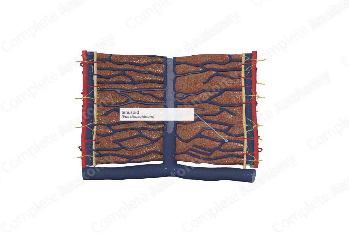
The sinusoids are blood capillaries that receive blood from the interlobular veins and arteries and carry it through the hepatic lobule parenchyma to the central vein. The sinusoids arising from the interlobular arteries join the neighboring sinusoids arising from the interlobular veins (which are terminal branches of the hepatic portal vein), which may take place after the sinusoids have traversed the hepatic lobule by a diameter of only one liver cell. The sinusoids extend from the portal triads in a “wave like” fashion throughout the lobule.
The sinusoids are lined with a thin and highly fenestrated endothelium with a discontinuous basal lamina. Unlike other blood capillaries, the sinusoids of the liver are also lined with specialized macrophages known as stellate sinusoidal macrophages, or Kupffer cells. The Kupffer cells are isolated in that they do not form junctions with their neighboring endothelium.
As the blood flows through the sinusoids, a substantial amount of the plasma is filtered, via the endothelial fenestrations, into a space between the sinusoid and the surrounding hepatocytes. This space is known as the perisinusoidal space (of Disse) and is typically 0.2-0.5 μm wide (Standring, 2016). The surfaces of the hepatocytes facing the perisinusoidal space have numerous microvilli, which creates a larger surface area of the hepatocyte exposed to the plasma leaking from the sinusoids. Hepatic stellate cells, or Ito cells, are also found in the perisinusoidal space.
Function
The sinusoids receive a mix of arterial (oxygen rich) and portal venous (nutrient rich) blood from the interlobular arteries and interlobular veins, respectively. These sinusoids pass through the parenchyma of the liver lobule and empty into the central vein. Along its course, plasma leaking into the perisinusoidal space is exposed to the basal surfaces of the hepatocytes, where exchange of materials between the plasma and hepatocytes can occur.
The sinusoids have a role in detoxifying the blood and in generating an immune response. This occurs via the Kupffer cells, which remove microbial and cellular fragments from circulation, as well as secreting cytokines into the blood. Kupffer cells are also primarily responsible for the removal of damaged red blood cells.
Ito cells are the primary storage site for vitamin A. They release retinol into the perisinusoidal space so that it is transferred to the retina in the blood. However, in pathological conditions, they differentiate into myofibroblasts and synthesize collagen.
List of Clinical Correlates
- Hemochromatosis
- Night blindness
References
Dorland, W. (2011) Dorland's Illustrated Medical Dictionary. 32nd edn. Philadelphia, USA: Elsevier Saunders.
Standring, S. (2016) Gray's Anatomy: The Anatomical Basis of Clinical Practice. Gray's Anatomy Series 41st edn.: Elsevier Limited.
