Quick Facts
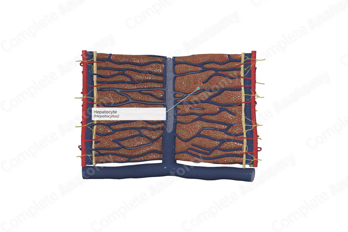
A hepatocyte is one of the polyhedral epithelial cells that constitute the substance of a liver acinus (Dorland, 2011).
Structure/Morphology
Hepatocytes are the main epithelial cells of the liver parenchyma. They make up around 80% of the liver volume and 60% of the liver cell population (Standring, 2016). They are polygonal in shape and have six sides but may have as many as 12 sides per cell. The hepatocytes are arranged as hepatic plates, which distribute outwards as radial cords from the central vein.
Hepatocytes have a large nucleus, spherical in shape, and located in the center of the cell. In the adult, many hepatocytes are binucleate and therefore are tetraploid (i.e., contain 4d amount of DNA). Two or more nucleoli are visible in each nucleus and heterochromatin is scattered almost evenly under the nuclear envelope (Mescher, 2013).
Hepatocytes are long-lived cells with an average lifespan of about five months. They are capable of considerable regeneration.
The cytoplasm of the hepatocytes has plentiful agranular (smooth) and granular (rough) endoplasmic reticulum, Golgi complexes, peroxisomes, and mitochondria, indicative of their high metabolic activity. Additionally, glycogen deposits and lipid droplets can be identified in the cytoplasm of hepatocytes.
Anatomical Relations
The basal surfaces of the hepatocytes face into the perisinusoidal space (of Disse) located adjacent to the sinusoids. These surfaces have numerous microvilli, which creates a large surface area that is exposed to the plasma that escapes from the sinusoids into this space.
The apical, or lateral, surfaces of adjacent hepatocytes are joined together via gap junctions and desmosomes. Localized intercellular spaces exist between the adjacent hepatocytes and are known as the bile canaliculi. Bile is secreted from the apical surfaces of the hepatocytes into the bile canaliculi. The tight junctions surrounding the canaliculi prevents bile from entering the sinusoids, thus creating a blood-bile barrier.
Function
Hepatocytes are the chief functional cells of the liver and are capable of completing metabolic, endocrine, and exocrine functions. The Golgi apparatus is involved in exocrine secretion of bile into the bile canaliculi, as well as synthesizing and secreting very low-density lipoproteins into the circulation (endocrine function).
The agranular (smooth) endoplasmic reticulum contains enzymes that are involved in the degradation of toxins and drugs and the synthesis of cholesterol. Lysosomes store iron in the hepatocytes, while the peroxisomes are involved in the detoxification of alcohol, breaking down fatty acids, and gluconeogenesis (production of glucose) (Pawlina, 2016).
Glycogen is also stored in hepatocytes, where abundant amounts are seen after a meal, while minimal amounts are seen after prolonged fasting.
List of Clinical Correlates
- Hemochromatosis
References
Dorland, W. (2011) Dorland's Illustrated Medical Dictionary. 32nd edn. Philadelphia, USA: Elsevier Saunders.
Mescher, A. (2013) Junqueira's Basic Histology: Text and Atlas. 13th edn.: McGraw-Hill Education.
Pawlina, W. 2016. Histology: A text and atlas with correlated cell and molecular biology. 7th ed. Philadelphia: Wolters Kluwer.
Standring, S. (2016) Gray's Anatomy: The Anatomical Basis of Clinical Practice. Gray's Anatomy Series 41st edn.: Elsevier Limited.
