Quick Facts
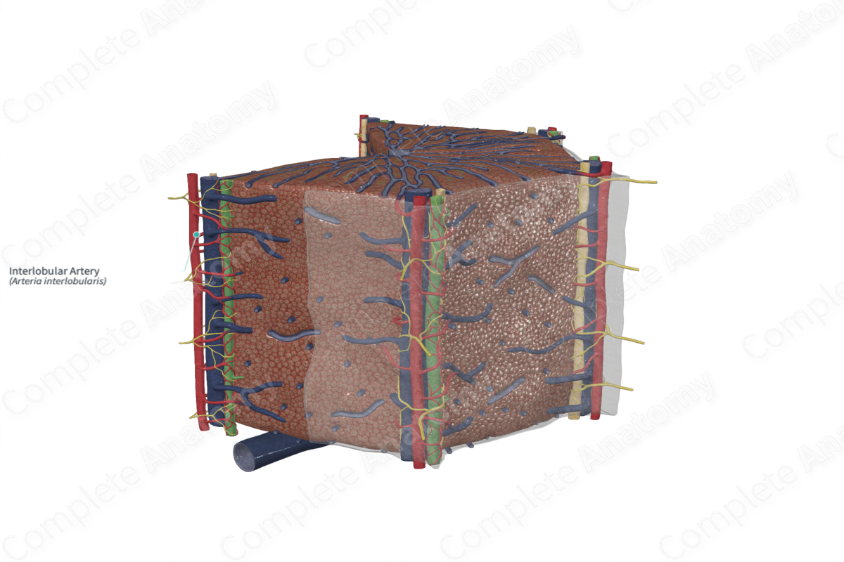
The interlobular arteries of liver are the arteries originating from the right or left branch of the hepatic artery proper, forming a plexus outside each hepatic lobule and supplying the walls of the interlobular veins and the accompanying bile ducts (Dorland, 2011).
Related parts of the anatomy
Structure/Morphology
The portal triad is the term given to the branches of the portal vein, hepatic artery, and bile duct that pass through the liver together. The term portal triad, however, is a misnomer, since lymphatic vessels and nerves also travel in this arrangement. The hepatic artery carries 25% of the total circulation to the liver, while the portal vein contributes to 75% of the total blood volume (Boron and Boulpaep, 2003). The blood of the hepatic artery is rich in oxygen as it has come directly from the celiac trunk of the abdominal aorta. The portal vein carries blood, rich not only in nutrients but also contains toxins, absorbed from the gastrointestinal tract.
The hepatic artery enters the liver at the porta hepatis, accompanied by the hepatic portal vein. The common bile duct and lymphatics exits the liver via the porta hepatis. The hepatic artery continues to bifurcate into segmental branches to each of the functional segments of the liver. Its terminal branches are the interlobular arteries, at the portal canals at the periphery of the hepatic lobule and send blood to the sinusoids.
The interlobular arteries are muscular arteries and are much smaller than the interlobular veins. They provide capillary networks to supply the interlobular veins (vas vasorum), interlobular bile duct, and parenchyma of the portal tract. The capillary networks either drain into the venous capillaries or enter the sinusoids directly (Krstic, 2013).
Anatomical Relations
The interlobular arteries accompany the interlobular veins and the interlobular bile duct. These vessels run parallel to each other in the portal tracts and form the portal triad and send blood to the sinusoids. They are also associated with lymphatic vessels and nerves.
Function
The interlobular arteries provide oxygenated blood to the liver parenchyma, including the connective tissue of the portal tracts. They also form multiple dense plexus around the interlobular vein and the interlobular biliary duct.
List of Clinical Correlates
- Veno-occlusive disease
References
Dorland, W. (2011) Dorland's Illustrated Medical Dictionary. 32nd edn. Philadelphia, USA: Elsevier Saunders.
Krstic, R. V. (2013) Human Microscopic Anatomy: An Atlas for Students of Medicine and Biology. Springer Berlin Heidelberg.
