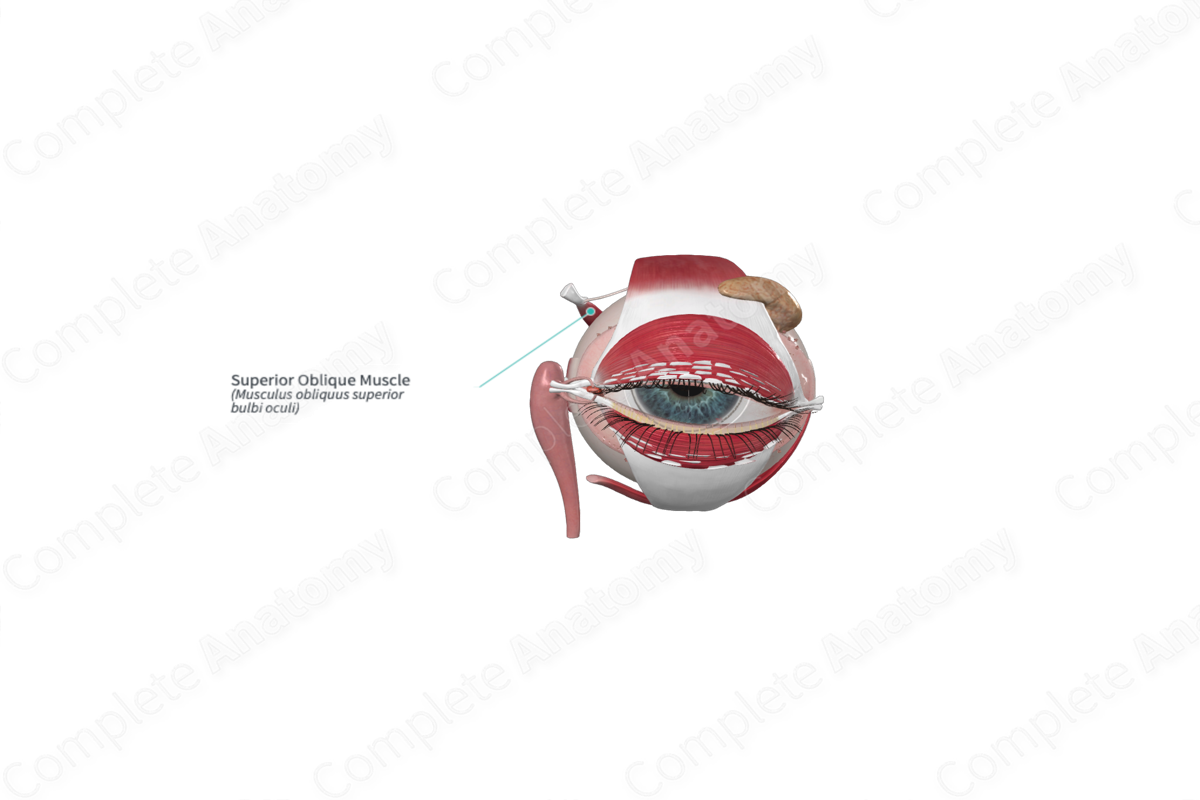
Superior Oblique Muscle
Musculus obliquus superior bulbi oculi
Read moreQuick Facts
Origin: Body of sphenoid bone.
Insertion: Sclera on the superolateral part of posterior quadrant of the eyeball.
Action: Depresses, abducts, and medially rotates eyeball.
Innervation: Trochlear nerve (CN IV).
Arterial Supply: Ophthalmic and supraorbital artery.


Origin
The superior oblique muscle arises from the body of the sphenoid bone, superomedial to the optic canal and the superior rectus muscle.
Insertion
From its origin, the superior oblique muscle traverses anteriorly in the superomedial corner of the orbit to reach a fibrous cartilaginous loop, the trochlea, in the anterior, superomedial corner of the orbit. Here, the muscle becomes tendinous. The tendon passes through the trochlea and passes posterolaterally to attach to the superolateral part of the sclera at the posterior quadrant of the eyeball.
Action
The superior oblique muscle directs the pupil inferiorly (depression) around the horizontal (transverse) axis, a movement that is strongest when the eye is adducted. Additionally, it rotates the eye laterally (abduction) around the vertical axis (strongest when the eye is already partly abducted by lateral rectus) and intorts the eye (medial rotation) around the anteroposterior axis.
List of Clinical Correlates
—Lesions of trochlear nerve
—Diplopia, especially when looking downwards as in walking downstairs, leading to falls.
