Quick Facts
Origin: Common tendinous ring.
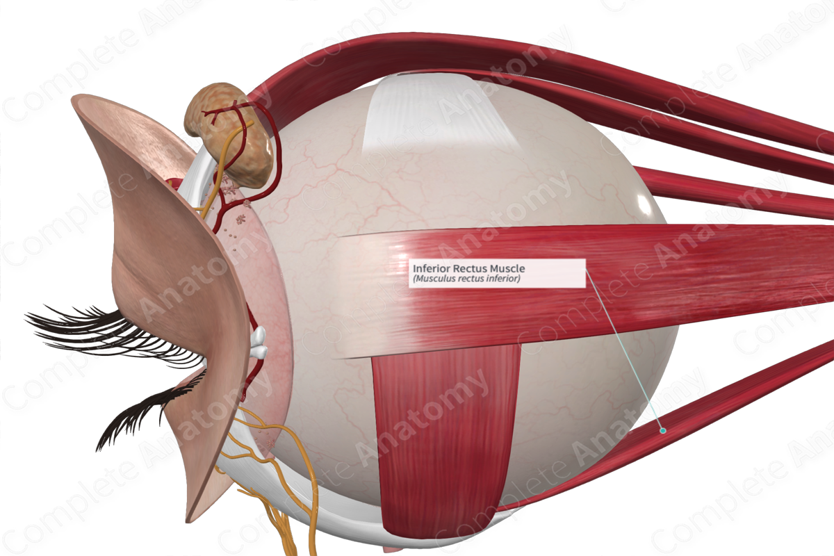
Insertion: Sclera, on inferior aspect of eyeball, just posterior to the corneosclearal junction.
Action: Depresses, adducts, and laterally rotates eyeball.
Innervation: Inferior branch of oculomotor nerve (CNIII).
Arterial Supply: Infraorbital artery; ophthalmic artery.
Origin
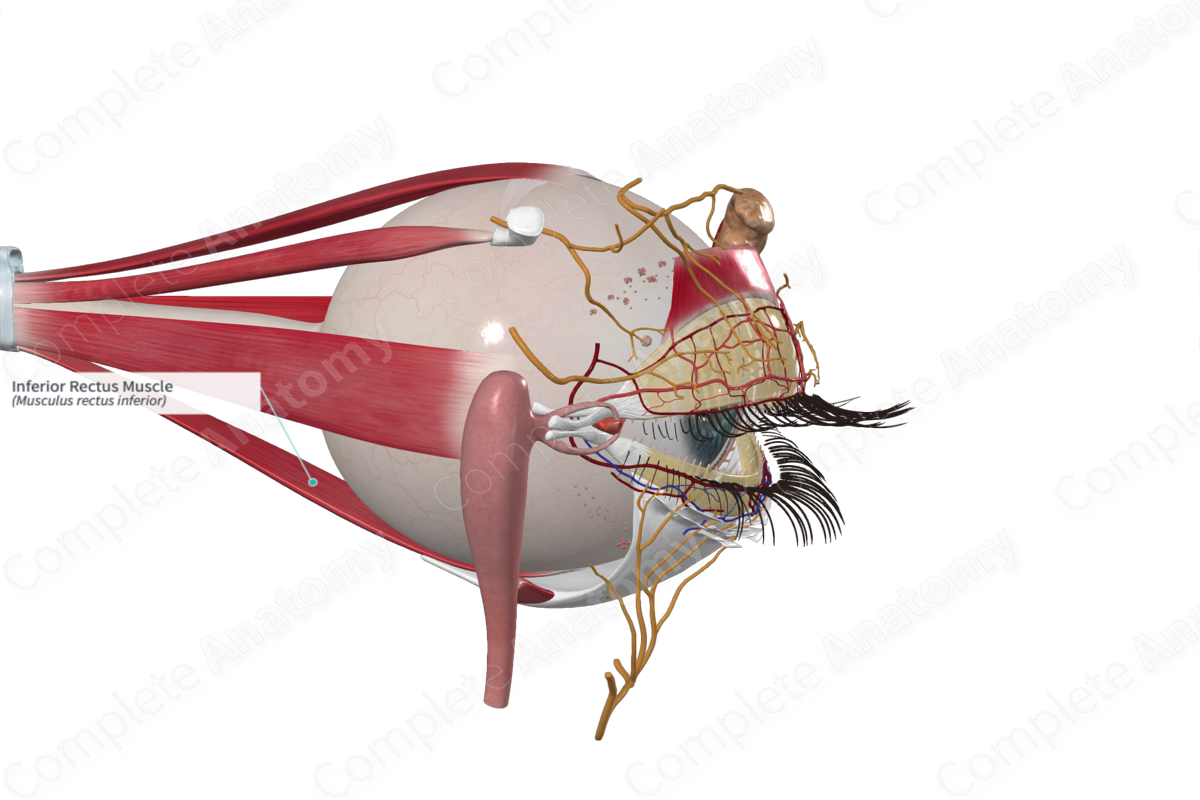
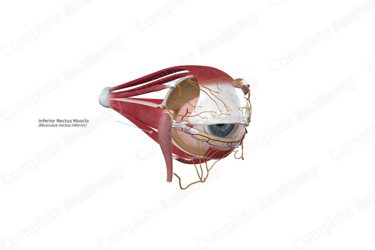
The inferior rectus muscle originates from a common tendinous ring, just beneath the optic canal.
Insertion
The inferior rectus muscle inserts on the sclera, obliquely, on the inferior aspect of the eyeball, posterior to the corneoscleral junction. Additional fibers extend from the inferior rectus muscle to the inferior tarsal plate of the lower eyelid. Therefore, when the eyeball is depressed by contraction of the inferior rectus muscle, i.e., gaze is directed downwards, the lower eyelid is also depressed.
Key Features & Anatomical Relations
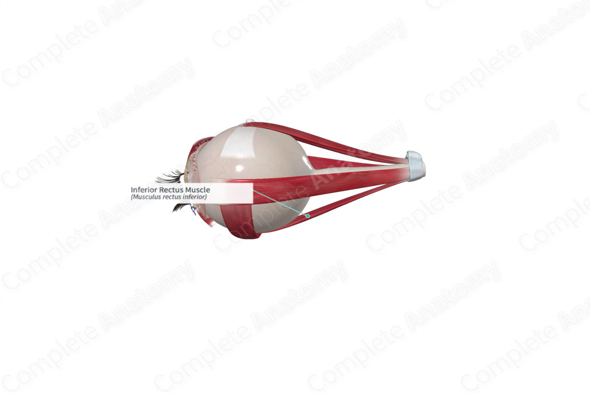
From the common tendinous ring, the inferior rectus muscle runs anterolaterally along the floor of the orbit.
Action
The inferior rectus muscle is primarily involved in depressing the eyeball around the transverse (horizontal) axis, i.e., moves the pupil inferiorly. It is most efficient at doing this when the eyeball is fully abducted. It is also involved in medial rotation (adduction) around the vertical axis and laterally rotation (extorsion) around the anteroposterior axis. These two actions are maximum when the eye is adducted.
