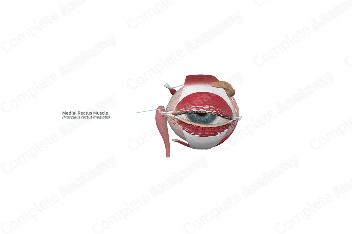
Quick Facts
Origin: Common tendinous ring.
Insertion: Medial surface of sclera.
Action: Adducts eyeball.
Innervation: Inferior branch of oculomotor nerve (CNIII).
Arterial Supply: Ophthalmic artery.


Origin
The medial rectus muscle arises from the upper and lower medial parts of the common tendinous ring. Additionally, some fibers of the medial rectus muscle arise from the dural sheath surrounding the optic nerve.
Insertion
From the common tendinous ring, the medial rectus muscle passes anteriorly in a horizontal plane, along the medial wall of the orbit. It is inferior to the superior oblique muscle. It inserts onto the medial surface of the sclera.
Key Features & Anatomical Relations
The medial rectus muscle is slightly shorter but strongest of the rectus group.
Action
The medial rectus muscle rotates the eyeball medially (adduction) around the vertical axis. When working in concert, the right and left medial recti muscles converge eyeballs so that the object of attention falls on bilateral maculae. Medial and lateral recti muscles work antagonistically producing saccadic and tracking motions.
List of Clinical Correlates
—Abducens nerve palsy
