Structure
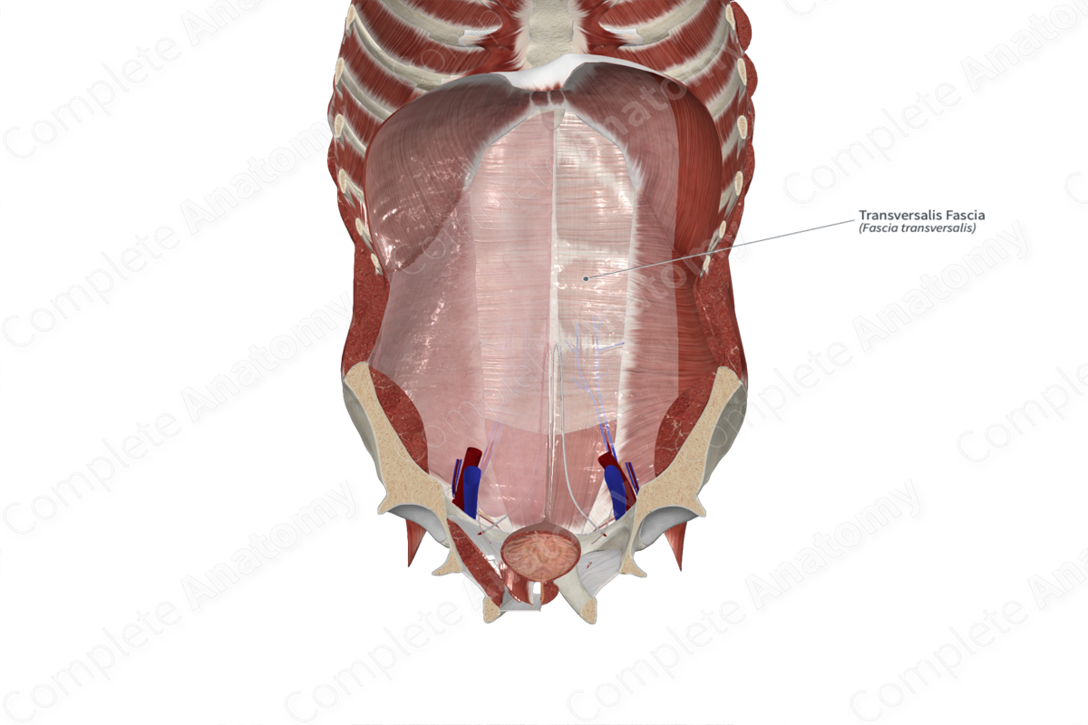
The transversalis fascia is an endoabdominal fascia that covers the internal surface of the transversus abdominis muscle. It is generally thin; however, it is thick and dense in the inguinal region.


Related parts of the anatomy
Anatomical Relations
The transversalis fascia sits between the transversus abdominis muscle and the extraperitoneal fat and peritoneum. It is continuous with the inferior surface of the diaphragm superiorly and the thoracolumbar fascia posteriorly. Inferiorly, the transversalis fascia merges with the iliac and pelvic fascia and attaches to the iliac crests at the posterior margin of the inguinal ligament, between the anterior superior iliac spine and the femoral sheath. It is also attached to the pubis by the conjoint tendon.
The iliopubic tract and interfoveolar ligament are inferior extensions and thickenings of the transversalis fascia. Posteriorly, the transversalis fascia blends with the thoracolumbar fascia.
The transversalis fascia extends through the deep inguinal ring to form part of the wall of the inguinal canal. The deep inguinal ring is an invagination of the transversalis fascia. It forms the roof of the lateral third of the inguinal canal and the posterior wall of the middle third of the inguinal canal.
The superior epigastric artery runs superiorly in the transversalis fascia, while the inferior epigastric artery pierces the transversalis fascia to enter the rectus sheath.
Function
The transversalis fascia separates the abdominal wall from the peritoneum. It covers the posterior aspect of the rectus abdominis muscles below the arcuate line. The transversalis fascia is also an important part of the wall of the inguinal canal, through which the round ligament (in females), and the vas deferens (in males) travel.
List of Clinical Correlates
—Direct inguinal hernia
—Preperitoneal hernia
Learn more about this topic from other Elsevier products

