
Unfortunately we don't fully support your browser. If you have the option to, please upgrade to a newer version or use Mozilla Firefox, Microsoft Edge, Google Chrome, or Safari 14 or newer. If you are unable to, and need support, please send us your feedback.

Elsevier
Publish with us
Diaphragmatic Fascia
Connective Tissue

Diaphragmatic Fascia

Fascia diaphragmatica

Read more

Structure

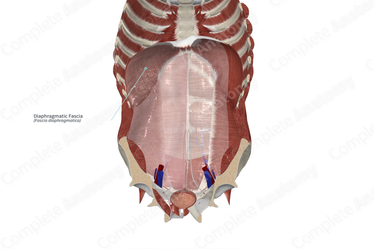
The diaphragmatic fascia is part of the endoabdominal fascia that lines the inferior surface of the diaphragm. It is continuous inferiorly with the transversalis and iliopsoas fascia.

Complete Anatomy
The world's most advanced 3D anatomy platform
Try it for Free

Anatomical Relations

Around the esophageal hiatus, the diaphragmatic fascia gives rise to the inferior phrenicoesophageal ligament, a thin extension of the diaphragmatic fascia that attaches to the esophagus.

Learn more about this topic from other Elsevier products

Fascia

ScienceDirect image

The fascia is also defined as a “the fascial system consists of the three-dimensional continuum of soft, collagen containing, loose and dense fibrous connective tissues that permeate the body” by Fascia Nomenclature Committee (Bordoni and Whitte, 2018), an as “masses of connective tissue large enough to be visible to the unaided eye” by Gray’s Anatomy (Standring, 2016).

Explore on ScienceDirect(opens in new tab/window)

Complete Anatomy

The world's most advanced 3D anatomy platform

Complete Anatomy