Quick Facts
Origin: Union of tributaries at the lateral angle of the orbit.
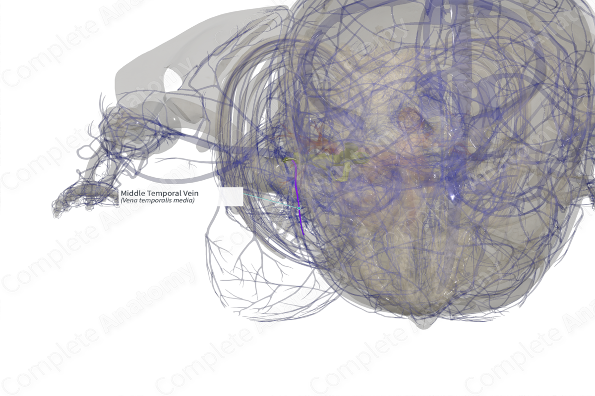
Course: Pierces the temporal fascia and passes backwards between the two layers of the deep temporal fascia to drain into the superficial temporal vein.
Tributaries: Zygomaticotemporal vein.
Drainage: Temporalis muscle.


Related parts of the anatomy
Origin
The middle temporal vein originates from two to four tributaries at the lateral orbital angle.
Course
The middle temporal vein passes backwards, downwards, and outwards in the temple region. It pierces the temporal fascia, then passes back between the superficial and deep layers of the deep temporal fascia, while being embedded within the superficial temporal fat pad (horizontal segment). As the vein courses posteriorly within the temporal fat pad, it makes a large curve downward in front of the ear and drains into superficial temporal vein, before it enters the parotid gland.
Tributaries
The middle temporal vein receives the zygomaticotemporal vein.
Structures Drained
The middle temporal vein drains blood from the temporalis muscle and the tissue of the temporal region.
Learn more about this topic from other Elsevier products

