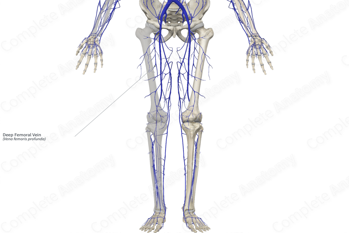
Quick Facts
Origin: Popliteal region.
Course: Travels between adductor muscles to join the femoral vein in the anterior compartment of the thigh.
Tributaries: Inferior gluteal, medial and lateral circumflex femoral veins, and muscular branches.
Drainage: Adductor, extensor, and flexor muscles of the thigh.
Related parts of the anatomy
Origin
The deep femoral vein is one of the main veins of the thigh. It originates distally in the popliteal region.
Course
The deep femoral vein runs posterior to the deep femoral artery as it traverses the entire length of the thigh. It joins the femoral vein approximately 3 cm distal to the inguinal ligament (Standring, 2016). Along its course, the vein passes between the adductor muscles and anastomoses with the popliteal vein.
Tributaries
Proximally, the medial and lateral circumflex femoral veins drain into the deep femoral vein, just inferior to the inguinal ligament. Distally, muscular branches join the vein along its course in the thigh.
The deep femoral vein anastomoses proximally with the inferior gluteal veins and distally with the popliteal vein.
Structures Drained
The deep femoral vein drains the adductor magnus, adductor longus, adductor brevis, pectineus, quadratus femoris, and iliopsoas muscles.
References
Standring, S. (2016) Gray's Anatomy: The Anatomical Basis of Clinical Practice. Gray's Anatomy Series 41 edn.: Elsevier Limited.
Learn more about this topic from other Elsevier products


